


Non-surgical treatment of hepatic hydatid cysts is a procedure in which the parasite is inactivated using special solutions by inserting thin needles directly into the cyst under ultrasound guidance, without making a surgical incision. In this method, the patient’s abdominal area is not opened with a scalpel, and the integrity of the organ is completely preserved.
Also known as the percutaneous approach, this technique works directly on the target thanks to the millimetric precision provided by medical imaging devices. The harmful structure inside the cyst is inactivated in place.
This closed system, frequently used in interventional radiology practice, completely eliminates long hospital stays and difficult recovery periods. After the procedure, only a mark the size of a needle puncture remains on the body.
This procedure, which is among the first options in current medical standards, maintains recovery comfort at the highest level. Individuals return to their social and professional lives quite quickly.
What Is Hepatic Hydatid Cyst Disease and How Is It Transmitted?
Hepatic hydatid cyst disease is caused by a microscopic parasite. The natural life cycle of this parasite generally takes place in the intestines of carnivorous animals such as dogs, wolves, and foxes. Parasite eggs released into the environment through animal feces are highly resistant to environmental conditions. These eggs, which spread to soil, drinking water, and agricultural areas through wind, rainwater, or insects, can remain viable for months. Humans become part of this cycle by consuming contaminated water from the environment, eating raw vegetables and fruits that have not been washed properly, or failing to maintain adequate hand hygiene after contact with animals carrying the parasite.
Parasite eggs entering the human body through the mouth resist stomach acid and travel directly to the small intestines. Upon reaching the intestinal environment, the thick outer shell of the egg opens, and the larval form emerging from it penetrates the intestinal wall and enters the body’s bloodstream. The vascular system in human anatomy carries blood absorbed from the intestines directly to the liver for purification and processing. Therefore, the vast majority of parasites traveling through the bloodstream become trapped in the narrow capillary network of the liver, which acts as a filter.
The parasite that settles in the liver tissue gradually begins to form a sac around itself to hide from the body’s immune system and survive. As this structure formed by the parasite expands by producing fluid from the inside outward, the human body also surrounds this foreign formation with connective tissue as a defense mechanism. This slowly growing formation, filled with fluid and covered externally by a protective layer produced by the body, is called a hydatid cyst. In most cases, the disease remains silent at this stage and continues to develop for a long time without causing any noticeable complaints.
What Symptoms Does Hepatic Hydatid Cyst Disease Cause?
A cyst located in the liver naturally exhibits an extremely slow growth rate. Its annual increase in diameter generally does not exceed a few centimeters. Such slow growth allows the disease to remain silent for many years without causing the patient any discomfort. In many cases, cysts are detected incidentally during ultrasound or tomography examinations performed for another health problem. However, as the cyst increases in volume and begins to press against the tense capsule surrounding the liver, various physical complaints start to appear.
The main complaints that may occur due to the pressure exerted by the cyst on surrounding tissues include:
- Pain in the upper right abdominal region
- Feeling of fullness in the stomach
- Early satiety
- Loss of appetite
- Difficulty digesting food
- Nausea
- Yellowing of the skin
- Yellowing of the whites of the eyes
- Darkening of urine color
- Fatigue
The severity of these symptoms varies depending on the size of the cyst, its location within the liver, and the degree of pressure it exerts on surrounding organs. When the cyst grows toward the stomach, early satiety and nausea become prominent, whereas pressure on the bile ducts slows the flow of bile into the intestine and causes findings such as jaundice. Pain felt in the upper right quadrant is generally described not as sharp or stabbing, but rather as a dull and continuous sensation of pressure.
What Cyst Characteristics Determine the Decision for Non-Surgical Treatment of Hepatic Hydatid Cyst Disease?
Not every fluid-filled sac detected in the liver has the same structural characteristics, and the course of the disease over time changes the internal architecture of the cyst. Therefore, before planning non-surgical treatment, the cyst must be evaluated and staged in detail using ultrasonography. Medical staging is used to determine the viability of the parasite and how suitable the cyst is for drainage.
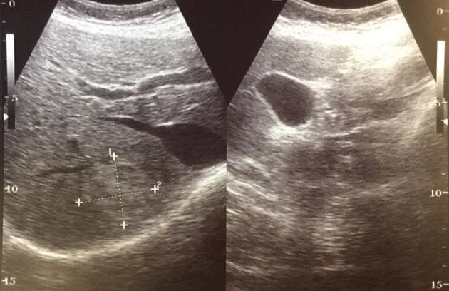
Early-stage cysts appear on the ultrasound screen as completely black, clear fluid-filled sacs. This indicates that the parasite is alive and active. Sometimes the inner membrane of the cyst gradually separates from the outer wall and begins to float in the fluid. Cysts in this active stage, with a clear internal structure or an inner membrane that has just begun to separate, are among the lesions that respond most favorably to non-surgical needle treatments.
As the disease progresses, the appearance of the cyst becomes more complex. New small daughter cysts may form inside the main cyst. This structure creates a multilocular, honeycomb-like appearance on ultrasonography. The fluid inside such complex cysts is generally dense and viscous, making it quite difficult to withdraw using standard needles. Therefore, honeycomb-like cysts require different non-surgical approaches involving larger-diameter tubes and irrigation of the cyst cavity.
In some cases, however, the immune system may have completely overcome the cyst. Over the years, the parasite loses its viability, its fluid dries up, and the cyst wall becomes calcified, turning into a hard scar. Since these calcified and inactive lesions are not expected to harm the patient, they are generally not treated with a needle; instead, they are monitored at regular intervals for any changes in size.
What Preparations Are Made for Patients Before Non-Surgical Treatment of Hepatic Hydatid Cysts?
To ensure that non-surgical procedures proceed comfortably and smoothly, there is a preparation phase that stabilizes the patient’s medical condition before the procedure. Oral antiparasitic medication is started approximately one week to ten days before the intervention to weaken the parasite’s reactions to external interventions. This medication penetrates the cyst and helps deplete the parasite’s energy resources and reduce its viability. This precaution greatly reduces the possibility of parasite remnants causing an unexpected reaction in the body during the procedure.
The medical preparation steps carried out on the day of the procedure include:
- Evaluation of blood tests
- Measurement of coagulation levels
- Establishment of intravenous access
- Administration of allergy-suppressing medications
- Monitoring of blood pressure and pulse values
- Monitoring of blood oxygen levels
- Administration of intravenous fluids for fluid balance
- Administration of sedative medications
During the preparation process, particular attention is paid to ensuring that the patient does not have a tendency to bleed. If any abnormalities are detected in laboratory values, corrective treatments are administered before the procedure. Because the fluid inside the cyst contains foreign proteins, medications containing cortisone or anti-allergy medications are administered intravenously to prevent a possible allergic reaction by the immune system. The patient remains awake in the procedure room, but the relaxing medications suppress feelings of fear and anxiety, allowing the procedure to be performed comfortably in a state between sleep and wakefulness.
How Is Non-Surgical Hepatic Hydatid Cyst Treatment with a Single Needle (PAIR) Performed?
The main method generally preferred for lesions smaller than six centimeters in diameter, consisting of a single cavity and containing no daughter cysts, is the PAIR technique. This method consists of inserting a needle into the cyst, withdrawing some of the fluid, administering therapeutic agents into the cyst, and finally draining everything inside.
At the beginning of the procedure, the ultrasound probe is moved over the patient’s skin to determine the point where the cyst can be seen most clearly and the safest approach angle can be achieved. Local anesthesia is administered at this point to numb the skin. As the needle advances, direct puncture of the cyst surface is avoided. Instead, the needle is guided through a certain distance of healthy liver tissue before reaching the cyst. Healthy liver tissue acts as a natural plug that prevents fluid from leaking out of the cyst. When the needle reaches the center of the cyst, some of the fluid is withdrawn with a syringe to reduce the internal pressure.
Once the pressure decreases, a medical contrast fluid is injected into the cyst through the needle, and the condition of the cyst is monitored in real time on the X-ray screen. The purpose of this contrast fluid is to check whether the cyst has a hidden connection with the surrounding bile ducts. When no leakage is observed, concentrated saline solution is introduced into the cyst to destroy the parasite and is left in place for a while. The purpose of the saline solution is to separate the inner membrane of the cyst from the main wall to which it is attached. This fluid is then withdrawn, and medical alcohol is injected to permanently dry out the cyst membrane. After the alcohol has remained in place for a sufficient period, it is completely aspirated, the cyst cavity is collapsed, and the needle is removed, completing the procedure in a single session.
How Does Non-Surgical Tube (Catheter) Treatment Work for Large Hepatic Hydatid Cysts?
When the cyst is larger than six centimeters or contains a very large volume of fluid, a single needle insertion is generally not sufficient to ensure that the cyst closes completely on the same day. Because the cyst is very large, when the fluid inside is suddenly drained, the enormous cavity left behind cannot immediately contract, and bodily fluids may accumulate in this space again. For this reason, in large lesions, the procedure is not completed by simply removing the needle; instead, a thin, soft, and flexible tube called a catheter is placed inside the cyst cavity.
The beginning of the procedure is the same as needle treatment. After the cyst is entered with a needle, a guidewire is passed through the needle. The needle is removed, and the flexible tube is advanced over this wire into the cyst. The end of the tube remaining inside the cyst has small holes and curls around itself to form a locking mechanism, preventing the tube from accidentally coming out. The other end remaining outside is connected to a drainage bag.
The fluid inside the large cyst is allowed to flow slowly and gradually into the bag. This gradual drainage is important to prevent sudden changes in pressure within the liver and damage to surrounding tissues. As the days pass, the cyst cavity slowly collapses and contracts. When the amount of fluid flowing from the tube into the bag decreases to a few drops and eventually stops, it indicates that the cyst cavity has narrowed considerably. At this point, medical alcohol is introduced through the tube to dry out the cyst and is withdrawn after remaining in place for a while. Once the procedure is completed, the tube is carefully removed, and the cyst treatment is finished.
How Is Non-Surgical Cleaning Performed in Complex Hepatic Hydatid Cyst Cases Containing Daughter Cysts?
In more advanced stages of the disease, the parasite continues to reproduce inside the cyst and forms hundreds of new small daughter cysts within the main cyst. A complex structure resembling overlapping circles or a honeycomb appears on the ultrasound screen. Between these daughter cysts, thick cyst sand with a jelly-like or muddy consistency accumulates. Because the contents are extremely dense and sticky, blockages occur when attempts are made to withdraw them using standard thin needles or narrow tubes.
To clean such complex cyst structures without surgery, special systems with designs different from standard methods are used. The access route extending from the abdominal wall to the liver is widened to accommodate larger-diameter, hose-like tubes that are resistant to blockages. After these wide-lumen devices are directed toward the center of the cyst, a mechanical irrigation process is initiated by introducing high-flow physiological saline.
With the help of aspiration devices and irrigation fluids advanced through the tube, the daughter cysts inside the main cyst are broken apart, sticky membranes are liquefied and mobilized, and the resulting mud-like remnants are forcefully aspirated. The inside of the cyst is cleared of all solid materials, as though it were being washed and cleaned with a vacuum. Once the cavity has been completely cleaned, the final stage is performed using drying medications to prevent fluid from being produced again. In this way, large lesions with complex structures can be treated.
What Non-Surgical Approach Is Followed When Hepatic Hydatid Cyst Fluid Leaks into the Bile Ducts?
The liver has an extremely extensive duct system that carries the bile it produces to the intestines. As a hydatid cyst grows within the liver, it may press against this duct system due to increasing internal pressure. This continuous contact and pressure may cause weakening between the cyst membrane and the bile duct, eventually resulting in the formation of a small connecting opening.
The presence of this connection is one of the most critical decision points in the treatment process. A yellowish-green color in the first fluid withdrawn with the needle or the flow of X-ray contrast material from the cyst into the surrounding bile ducts confirms the presence of such a leak. If a connection between the cyst and the bile ducts is detected, the administration of medical alcohol or other caustic agents into the cyst is avoided. If these medications leak into the bile ducts, there is a risk of damage and narrowing in the ducts.
When such a situation is encountered, the non-surgical treatment strategy is changed. Only a drainage tube is placed inside the cyst, allowing the fluid to flow out. When the excessive pressure inside the cyst decreases, the small opening in the bile duct often closes due to the liver’s natural elasticity. If the leakage does not stop, an endoscopic camera technique advanced through the stomach and small intestine is used to widen the point where the main bile duct empties into the intestine. This procedure allows bile to flow easily into the intestine instead of leaking into the cyst, creating favorable conditions for the opening to heal. Once the leakage stops, the drying procedure can safely continue.
Do the Medications Used During Non-Surgical Hepatic Hydatid Cyst Treatment Have Side Effects?
The main factor contributing to success in percutaneous treatment is the destructive effect of the medical solutions applied inside the cyst on the parasite membrane. While concentrated saline solution (hypertonic saline) disrupts the cellular structure, medical alcohol (ethanol) chemically dries out the cyst wall. Since these substances have powerful effects, it is extremely important that they remain only within the boundaries of the targeted cyst.
If the chemicals used leak outside the cyst, for example into the abdominal cavity, they have the potential to cause local pain or regional irritation. Similarly, if excessive amounts of highly concentrated saline solution are used and enter the systemic bloodstream uncontrollably, changes in the body’s sodium balance may be observed. However, the detailed medical protocols applied are designed to minimize the possibility of such side effects.
Before these solutions are administered, the internal volume of the cyst is carefully calculated using ultrasound images. The amount of chemical solution introduced is always kept lower than the amount of fluid withdrawn from the cyst. This rule prevents an increase in pressure inside the cyst and largely prevents the chemical from leaking out. After the administered agents remain inside the cyst only for the intended duration, they are completely withdrawn using a syringe. Since no medical chemicals are left in the body after the procedure, the aim is to avoid unwanted side effects.
What Are the Advantages of Non-Surgical Hepatic Hydatid Cyst Treatment Methods Compared with Traditional Open Surgery?
In previous years, the main treatment for hepatic hydatid cyst disease was based on cleaning the cyst through large abdominal incisions or removing part of the liver together with the cyst. Needle- and catheter-based methods, which have become more widespread with advancing technology, offer many clinical benefits that improve patient comfort during recovery. Since no large surgical wound is created in the body, tissue trauma remains very limited, and many of the difficulties observed after surgery are avoided.
The main advantages offered to patients by non-surgical methods include:
- No surgical incision on the skin
- Low risk of incision-related infection
- No need for general anesthesia
- Preservation of healthy liver tissue
- Low levels of post-procedure pain
- Very short hospital stay
- Elimination of the possibility of incisional hernia formation in later years
- Rapid return to daily and professional life
While surgical methods involve general anesthesia, which requires the patient to be placed into a deep sleep, non-surgical procedures involve numbing only the needle insertion site and administering relaxing medications intravenously. The patient breathes independently during the procedure. Compared with hospital stays lasting several days, patients undergoing needle-based treatments can generally be discharged on the same day or after one day of observation and continue their social lives. Since the cyst fluid is drained using needles within a closed system, the risk of the parasite spreading to the peritoneum is also significantly reduced compared with surgery.
How Is the Recovery Process Monitored After Non-Surgical Hepatic Hydatid Cyst Treatment?
After the procedure performed with a needle or catheter is completed, the cyst does not immediately disappear from the ultrasound screen. The primary aim of the treatment is not to remove a piece of the liver, but to inactivate the parasite and collapse the tense, fluid-filled structure of the cyst. Over time, the dried cyst wall wrinkles, collapses, and folds in on itself, turning into harmless scar tissue. This shrinkage and healing process extends over several months.
A specific follow-up program is implemented to ensure that recovery progresses properly. Patients are generally advised to continue oral medication for a while during the first weeks after treatment to suppress possible microscopic parasites that may circulate in the blood. At the first follow-up examination one month after the procedure, blood tests showing liver enzyme levels are evaluated. The presence of conditions such as infection or fluid accumulation in the early period is examined.
Subsequent follow-ups are generally performed at three- or six-month intervals during the first year using ultrasonography. During these examinations, it is observed that the cyst decreases in size, the fluid inside completely disappears, and the tissue gradually becomes more solid. Over time, the area surrounding the former cyst becomes increasingly calcified and is isolated by the body. The absence of renewed fluid production or an increase in cyst size indicates that the parasite has lost its viability and the lesion has become completely harmless. After the first year, patients are invited for standard ultrasonographic examinations once a year as a precaution, allowing their condition to continue to be monitored safely.











Vaka Örnekleri