


Hemorrhage is a serious medical condition that occurs when blood leaks out of the vessels due to the disruption of vascular integrity. It may develop due to trauma, surgical procedures, vascular diseases, or clotting disorders. Early diagnosis and rapid medical intervention are critically important in preventing vital organ damage and life-threatening complications.
Symptoms of internal bleeding may present with different clinical findings depending on the location and severity of the bleeding. Dizziness, weakness, rapid pulse, low blood pressure, and changes in consciousness are among the most common symptoms. Severe internal bleeding can impair organ functions and create a life-threatening risk that requires emergency intervention.
Types of hemorrhage are classified according to the anatomical location and source of the bleeding. Arterial, venous, and capillary bleeding are categorized based on the type of vessel, while internal hemorrhage and external hemorrhage are defined according to whether the bleeding occurs inside or outside the body. This classification plays an important role in determining the appropriate treatment approach.
Hemorrhage treatment methods include medical interventions planned according to the cause, location, and severity of the bleeding. While simple bleeding may be managed with compression and fluid support, serious cases may require surgical intervention, blood transfusion, and coagulation therapies. Fast and accurate treatment significantly reduces the risk of complications.
| Things You Should Know | Information |
| Definition | Hemorrhage is the leakage of blood outside the vessels due to the rupture or damage of blood vessels within the body. It can occur as internal or external bleeding. |
| General Classification | It is divided into two main groups: Internal Hemorrhage (bleeding occurring within the body) and External Hemorrhage (bleeding flowing out of the body). |
| Causes | Trauma (accidents, falls), surgical complications, ulcers, hypertension, aneurysms, tears during childbirth, bleeding disorders (hemophilia), tumors. |
| Symptoms | Sudden or gradual blood loss, weakness, pallor, dizziness, rapid pulse, low blood pressure, cold sweating, loss of consciousness. In internal bleeding, abdominal pain, swelling, and shortness of breath may also be observed. |
| Types (According to Source) | Arterial Bleeding (bright red, rapid), Venous Bleeding (dark red, continuous), Capillary Bleeding (superficial, slow). |
| Types (According to Location) | Gastrointestinal Bleeding, Intracranial Bleeding, Pulmonary Bleeding, Urogenital Bleeding, Traumatic Bleeding, Postpartum Hemorrhage. |
| Diagnostic Methods | Physical examination, blood tests (hemoglobin/hematocrit), ultrasound, CT (Computed Tomography), MRI, endoscopy, angiography. |
| Emergency Intervention | Stopping the bleeding (compression, tourniquet), fluid therapy, oxygen support, blood transfusion, rapid surgical intervention. |
| Medical Treatment | Ligation of the bleeding vessel, endoscopic intervention, embolization, surgical resection, drug therapy (for example clotting agents or tranexamic acid). |
| Follow-up and Monitoring | Monitoring vital signs, tracking hematological values, preventing recurrent bleeding, treating the underlying cause. |
| Complications | Shock, organ failure, anemia, hypovolemic shock, risk of death (depending on the severity of bleeding and intervention time). |
| Preventive Measures | Regulation of anticoagulant use, control of high blood pressure, avoiding trauma, regular health check-ups. |
What Is Hemorrhage (Bleeding) and What Process Does It Initiate in Our Body?
Imagine a vast transportation network that surrounds our body from head to toe, carrying oxygen and nutrients that give life to our organs. This network consists of our blood vessels. Hemorrhage, in its simplest and most understandable definition, is the leakage of blood outside the vessel as a result of damage occurring at any point in this vital transportation network. The escape of blood outside the vessel does not only mean a physical fluid loss; it also means a severe disruption of the body’s perfect internal balance.
When vascular integrity is disrupted and blood begins to leave the vessel, the total fluid volume in our body suddenly decreases. The heart, like a pump with decreasing water, begins to fail in sending sufficient blood to the organs. As soon as our body detects this situation, it activates an extraordinary defense mechanism. Alarm bells ring, the nervous system is stimulated, and the heart starts beating much faster in hopes of pumping more blood. At the same time, vessels in the skin surface and non-vital areas constrict and narrow; the aim is to direct the limited remaining blood to the most critical organs such as the brain and heart. The main reason people suddenly become pale and sweat coldly during bleeding is this protective mechanism.
However, if the bleeding cannot be controlled, this tremendous defense system becomes exhausted and helpless after a while. Tissues and organs that cannot receive blood begin to suffer from oxygen deprivation. Cells deprived of oxygen try to produce energy without oxygen by abandoning their normal functioning systems in order to survive. This leads to the accumulation of harmful substances such as lactic acid in tissues, which causes fatigue in muscles. If this chain of damage is not broken by external intervention, cells begin to die and irreversible organ failures occur.
What Are the Types of Hemorrhage According to Vascular Structure?
Our vascular system does not consist of a single type of structure. We have vessels with different functions and structures that carry blood from the heart, to the tissues, and back to the heart. Therefore, bleeding also shows very different characteristic features depending on the type of vessel it originates from. These features are extremely important to understand the urgency of the situation.
The main sources of bleeding are:
- Arteries
- Veins
- Capillaries
Arteries are the main routes that carry fresh and oxygen-rich blood pumped from the heart to the body. Blood in these vessels moves with very high pressure with each heartbeat. Therefore, arterial bleeding is bright red in color and flows in spurts in synchronization with the heartbeat. Since they can lead to an incredible amount of blood loss in a very short time, they are the most life-threatening type of bleeding and require intervention within seconds.
Veins, on the other hand, are return routes that carry used and oxygen-depleted blood from the tissues back to the heart and lungs to be cleaned. The pressure in veins is much lower than in arteries. The color of the blood is darker, closer to burgundy, because it has less oxygen. The bleeding does not spurt but occurs as a continuous seepage or flow. Although it does not create a shock picture within seconds like arterial bleeding, very insidious and fatal blood losses may occur especially if large veins in the abdomen or chest cavity are damaged.
Capillaries are the smallest and finest branches of this system. They are microscopic vessels where substance exchange with cells occurs. Capillary bleeding is usually seen in minor abrasions, paper cuts, or superficial injuries encountered in daily life. The bleeding occurs drop by drop or as a slight seepage and usually stops on its own in a short time thanks to the body’s natural clotting mechanism.
What Are the Main Differences Between Internal and External Hemorrhage?
Where bleeding occurs in the body is another important factor that completely changes the diagnosis and treatment process. External bleeding is the situation where blood flows directly out of the body through a wound or cut where the integrity of the skin is disrupted. Since it can be clearly seen with the naked eye, it is relatively easier to notice and apply first aid by applying pressure.
Internal bleeding, on the other hand, occurs entirely within the closed compartments of the body. Blood does not exit the body; instead, it leaks into the chest cavity, abdominal cavity, joint spaces, muscles, or directly into the tissues of organs such as the brain, liver, and spleen. The greatest danger of internal bleeding is that it is “insidious.” Since no blood is seen externally, the seriousness of the situation is often recognized late. In order to diagnose such bleeding, changes in the patient’s general condition must be observed very carefully, a strong suspicion must be maintained, and the inside of the body must be visualized with technological devices without losing time.
What Are the Symptoms of Internal Hemorrhage in Different Regions of the Body?
Although internal bleeding is not visible to the eye, it gives very diverse and sometimes very specific signs depending on the area where it accumulates and the pressure it exerts on organs. Common alarms that occur due to a decrease in blood volume throughout the body should always be taken into consideration.
General and common internal bleeding symptoms are:
- Pallor
- Sweating
- Tachycardia
- Weakness
- Thirst
- Dizziness
In addition to these general symptoms, our body sends different SOS signals depending on the specific region where the bleeding occurs. For example, in intracranial (cerebral) hemorrhages, due to the closed and non-flexible structure of the skull, the accumulated blood puts serious pressure on the brain. As a result of this pressure, a sudden and extremely severe headache that has never been experienced before occurs. This is usually accompanied by sudden projectile vomiting without nausea. Visual disturbances, inequality in pupil sizes, speech impairment, tingling on one side of the body, or complete paralysis are the most typical findings of brain hemorrhages.
In chest cavity and lung bleeding, the main problem is difficulty in breathing. When blood accumulates around or inside the lungs, the lungs cannot expand sufficiently. The patient cannot take a deep breath, feels a stabbing pain in the chest, and may experience a sensation of suffocation. Additionally, fresh, sometimes foamy blood may come from the mouth with severe coughing attacks.
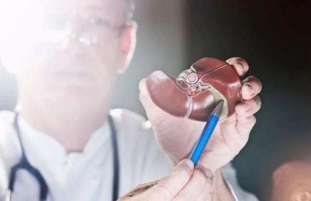
Intra-abdominal bleeding usually occurs due to damage to the liver or spleen after severe impacts such as traffic accidents or falls from height. A very severe sensitivity begins in the abdominal area upon touch. The abdominal muscles contract reflexively to protect the damage inside, and the abdomen becomes hard like a board. In the following hours, bruising under the skin around the navel or on the sides of the waist may be noticed.
In bleeding in the stomach and intestinal system, the situation is usually noticed through blood expelled from the digestive tract. In stomach bleeding, the person either vomits fresh red blood or vomits clotted material resembling coffee grounds because the blood is digested in the stomach. If the blood moves downward through the intestines, the stool becomes tar-like, pitch black, sticky, and very foul-smelling. In bleeding involving the urinary tract and kidneys, the color of the urine turns pink, tea-colored, or directly blood red.
What Are the Stages of Hemorrhagic Shock Due to Blood Loss?
In medical literature, a system called “hemorrhagic shock stages” is used to understand the destructive effects of bleeding on the human body and to classify the urgency of the patient’s condition. These stages are determined according to how much of the total blood in the body is lost and how the organs respond to this situation. There are four main stages.
Shock stages are:
- First
- Second
- Third
- Fourth
The first stage is the mildest form of shock. Less than fifteen percent of the total blood in the body has been lost. For example, when we donate blood, we actually experience a very mild blood loss close to the first stage, and our body tolerates it within seconds. In this stage, blood pressure is normal, and the patient’s consciousness is completely intact. There may be a very slight increase in heart rate that the person may not even notice.
In the second stage, blood loss has reached between fifteen and thirty percent. At this point, the body begins to struggle. The heart starts beating much faster to compensate for the decreased blood volume. The patient breathes faster as if out of breath. Blood pressure is generally still within normal limits, but the value we call pulse pressure begins to narrow. The patient feels restless, slightly frightened, and weak. A decrease in urine output is observed because the blood going to the kidneys is reduced.
The third stage is a severe stage of shock where alarm bells are ringing loudly. Blood loss has reached the thirty to forty percent range. The body’s ability to compensate has now failed. Significant and dangerous drops are detected in blood pressure measurements. Since sufficient oxygen does not reach the brain, confusion begins in the patient’s consciousness. The person may give meaningless answers to questions, become extremely irritable or, on the contrary, completely unresponsive to the environment. The skin is ice cold, and the survival of organs is hanging by a thread.
The fourth stage represents a critical line between life and death. More than forty percent of the blood has left the body. The pulse becomes so weak that it cannot be felt from the wrist. The skin is not only cold but also takes on a mottled purplish appearance like marble. The kidneys have completely stopped functioning, so there is no urine output at all. The patient is usually in a deep coma. In this stage, even minutes or seconds are crucial; if not intervened immediately, the situation unfortunately results in death.
What Are the Modern Imaging Methods Used in the Diagnosis of Hemorrhage?
Finding the location of internal bleeding is like searching for a needle in a dark room. To find these leaks that we cannot see with the naked eye, we need advanced imaging technologies, which are the eyes of modern medicine. When a patient arrives at the hospital with suspicion of trauma or internal bleeding, the process progresses very quickly.
The main imaging methods used are:
- X-ray
- Ultrasonography
- Tomography
- Angiography
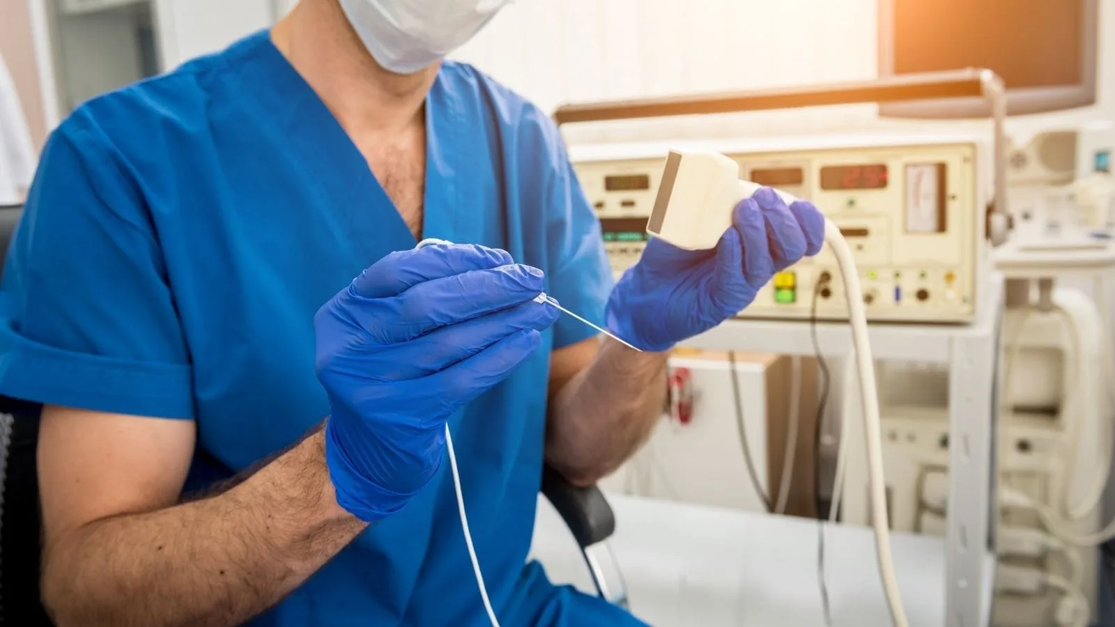
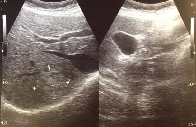
Simple X-ray films are mostly used to detect bone fractures. Ultrasonography devices are a very fast and practical method used in emergency departments at the patient’s bedside to understand within seconds whether fluid (blood) has accumulated in the abdomen. However, the main detailed examination is performed with Computed Tomography (CT) devices. With the help of a special dye administered intravenously, the inside of the body is mapped in three dimensions and with high resolution. Tomography clearly reveals around which organ the blood has accumulated and whether there is a rupture in the organs.
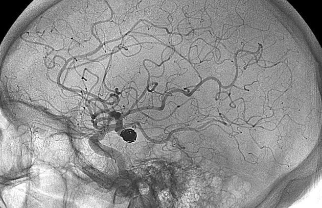
However, in order to see exactly from which vessel the bleeding is leaking and to treat it at the same time, “Angiography” is always the most definitive and advanced method. In the angiography procedure, a contrast agent is administered by entering the vessel, and the inside of the vascular network is monitored on the screen like a live video broadcast.
What Role Does Interventional Radiology Play in the Treatment of Hemorrhage?
In the past, the only way to stop an internal bleeding deep within the body was to open the patient’s abdomen or chest completely and find and ligate that vessel. These open surgeries carried enormous risks for a patient who had already lost a lot of blood and gone into shock. However, today, thanks to the branch of medicine equipped with high technology called “Interventional Radiology,” the rules of the game have completely changed.
Interventional radiology is the art of reaching the source of the problem by traveling “inside” the vessels instead of making large incisions from the outside. The procedure is extremely delicate and astonishing. A small incision the size of a needle hole is made from a vessel in the patient’s groin area or sometimes from the wrist. Through this tiny opening, very thin, soft, flexible plastic tubes (catheters) as thin as a hair strand are placed into the vessel.
Under the guidance of angiography images on large screens, these thin tubes are slowly advanced through the winding paths of the vessels. It is possible to reach every point the vascular system extends to, from the neck vessels to the deepest folds of the brain, from the abdominal aorta to the most distal branches of the intestine. The tip of the catheter is brought right into the bleeding and ruptured vessel. This approach is called “minimally invasive” in medicine, meaning it causes the least harm. The patient is usually awake during this procedure, there is no need for full general anesthesia, and the entire life-saving process is completed with only local anesthesia and mild sedatives.
What Are the Embolization Agents Used to Stop Hemorrhage?
After finding the bleeding vessel from the inside, the next step is to stop the blood by permanently or temporarily blocking that vessel. This procedure is called “Embolization.” Various technological materials are used to close the vessel from the inside; these materials are called embolic agents.
Commonly used materials are:
- Coils
- Particles
- Adhesives
- Sponges
Coils are microscopic wires made of platinum or various metals. When pushed into the vessel through thin tubes, they fold over themselves inside the vessel and take the form of a bundle or spring. This metal bundle physically blocks the blood flow and allows a clot to form quickly on it, permanently closing the vessel. They are frequently used especially in arterial bleeding and in the treatment of aneurysms in brain vessels.
Particles or microspheres are tiny beads the size of salt or sand grains. They are mostly used by injecting them in liquid form to block capillary bleeding spread over wide areas, tumor bleeding, or the capillary branches of the intestine. They get stuck at the narrowest point they reach and stop blood flow.
Liquid adhesives (special medical substances such as Glue or Onyx) work like the fast adhesives we know in daily life, but they are specially produced for medical use. The moment they are injected into the vessel, they solidify within seconds upon contact with blood and turn into a hard plastic mold. They act as an immediate plug in severe bleeding.
Sponge-like materials (Gelfoam) are used in cases where we do not want to completely destroy the vessel but only want to create a temporary blockage for a few weeks. The body dissolves this sponge over time, and the vessel reopens.
What Is Done in Stomach and Intestinal (Gastrointestinal) Hemorrhages?
Bleeding in the stomach and intestinal system is one of the most common problems handled in emergency departments. Bleeding from stomach ulcers, duodenal lesions, or esophageal varices caused by alcohol and certain diseases such as cirrhosis can be quite dangerous. In most cases, these bleedings are treated by entering the stomach with endoscopy devices equipped with cameras.
However, sometimes the bleeding is so severe that endoscopy cameras cannot see due to the presence of blood, or the interventions fail. At this point, interventional radiology comes into play and solves the problem from the inside, through the vascular route. Among the many vessels supplying the stomach or intestine, the bleeding vessel feeding the ulcer is identified and blocked internally with particles or coils.
In severe variceal bleeding due to liver cirrhosis, a much more specialized procedure is performed. This procedure is called TIPS. Because the liver becomes hardened, blood cannot pass through it and exerts excessive pressure on the delicate vessels in the esophagus, causing them to rupture and bleed severely. With interventional radiology techniques, access is gained through the neck vessel into the liver, and a special tube (stent) is placed between the two major vascular systems inside the liver. This creates a new and easier tunnel for blood flow, suddenly reducing the excessive pressure in the vessels and immediately stopping the variceal bleeding.
Which Methods Are Used in Severe Hemorrhages of Pulmonary Origin (Hemoptysis)?
The condition where more than half a glass of blood or continuous blood is coughed up from the mouth is called hemoptysis. It is usually caused by structural damage to the vessels in lung tissue due to past tuberculosis, lung infections, or lung tumors. The most frightening aspect of this bleeding is the risk of blood filling the patient’s airway and healthy lungs, leading to suffocation.
In such a critical situation, performing open lung surgery is extremely risky. Instead, patients are quickly taken to the angiography unit. By entering through the groin, the vessels supplying the lung and causing bleeding are identified. Microscopic particles are injected into these vessels to dry the bleeding from the inside. This is an incredibly life-saving and comfortable method applied without opening the chest cavity and without intubating the patient.
How Is Life Saved in Hemorrhages Caused by Traffic Accidents and Trauma?
Major traffic accidents, falls from height, or workplace accidents cause severe impacts on the body. Even if no external bleeding or wounds are visible, internal organs may rupture due to the impact. Massive internal bleeding occurs especially in cases of liver and spleen ruptures or fractures of the hip bones called the pelvis. The inside of the pelvis is woven with a very rich vascular network, and fracture ends can cut these vessels, causing liters of blood to accumulate within minutes in the back wall of the abdomen.
In modern trauma centers today, the approach is as follows: if the patient’s blood pressure is rapidly decreasing, long open surgeries will further weaken the patient’s resistance. Therefore, the patient is directly taken to the interventional radiology angiography suite. Within minutes, access is gained through the groin, and the vessels that are torn between the fractured bones and leaking blood are identified. These vessels are quickly blocked with coils or liquid adhesives, stopping the blood flow. Thus, the patient is stabilized, blood pressure rises, and the patient holds on to life.
How Is the Uterus Protected in Postpartum Hemorrhage?
The birth process, where a new life comes into the world, is miraculous, but sometimes it may involve unexpected complications. In both normal deliveries and cesarean sections, if the uterus fails to contract adequately (atony) after the baby is delivered, very severe and uncontrollable vaginal bleeding may occur. In the past, if the bleeding could not be stopped despite medications, the only and final option to save the mother’s life was to completely remove the uterus surgically. This meant that the mother could never have children again.
Today, medicine has a much more delicate and organ-preserving option. When the mother’s condition begins to deteriorate, a thin tube is inserted through the groin vessel and advanced directly to the arteries supplying the uterus. These vessels are blocked with temporarily dissolvable sponge-like materials (Gelfoam). The bleeding stops immediately, the mother recovers from shock, and most importantly, the uterus remains in place. When the body dissolves these temporary sponges within weeks, the vessels reopen, and the mother can become pregnant again in the future.
How Is Stent Treatment Applied in Hemorrhages Due to Aortic Ruptures?
The aorta is our main highway that carries blood from the heart and distributes it to the entire body. Sometimes, due to high blood pressure, aging, or genetic factors, weaknesses and bulging (aneurysm) develop in the wall of this massive vessel. If this bulging area suddenly ruptures or bursts one day, blood rapidly fills the abdominal or chest cavity within seconds. This is one of the most urgent and dramatic fatal scenarios in medicine.
In the past, it was necessary to completely open the chest or abdomen of patients in this condition and replace the ruptured vessel with an artificial tube. It was very difficult to withstand this surgery. Now, thanks to advanced interventional radiology techniques, intervention can be performed using a closed method by entering through the vessel in the groin. A fabric-covered, very strong wire mesh tube (stent-graft) is advanced through the vessel and placed exactly into the ruptured area. This prosthetic tube, which opens inside like an umbrella, seals the leakage from the inside by allowing blood to pass through a new tunnel.
What Are the Advantages of Interventional Radiology Compared to Open Surgery?
Interventional radiology has completely changed the vision of modern medicine. The principle of providing maximum benefit with minimal harm to the patient is the fundamental philosophy of this field. The benefits it provides compared to large, open surgeries are revolutionary for both the patient and the hospital.
The advantages are:
- No incision
- Fast recovery
- Less pain
- Low risk of infection
Since all procedures are performed through a hole of only a few millimeters, large wounds requiring sutures do not occur. This almost completely eliminates postoperative pain. Since there is no open wound, the risk of infection is almost negligible. Most patients undergo these procedures without heavy general anesthesia, only with sedatives that induce a state of drowsiness. The greatest advantage is the recovery time. Instead of staying in the hospital for days, most patients can get up the next day after the procedure, be discharged, and return to their normal daily lives within a few days.
What Should Be Considered During the Recovery Process After Hemorrhage Treatment?
As important as the success of the treatment is, the recovery period spent at home afterward is also of vital importance. No matter how small the intervention may seem, it should not be forgotten that the body has gone through a major crisis internally and has lost a significant amount of blood. It takes time for the lost blood cells to be replaced and for the organs to regain their previous performance.
The rules to be considered during this process are:
- Rest
- Plenty of fluid intake
- Regular nutrition
- Doctor follow-up
Patients should absolutely avoid heavy physical exercises, lifting heavy objects, and strenuous work during the first few weeks. Consuming plenty of water and eating foods rich in iron and vitamins that support blood production are very important to restore lost blood volume. The small wound dressing at the entry site should be kept clean, and it should be monitored for swelling or bruising. Most importantly, follow-up tomography or ultrasound scans scheduled by the doctor must not be neglected to monitor the success of the procedure.
Frequently Asked Questions
What Is Hemorrhage and how does it occur within the body?
Hemorrhage is the loss of blood from damaged blood vessels due to injury, surgery, disease, or vascular abnormalities. It can occur internally or externally and may range from mild bleeding to life-threatening blood loss.
What symptoms may indicate a serious hemorrhage?
Symptoms of significant hemorrhage can include dizziness, weakness, rapid heartbeat, low blood pressure, confusion, pale skin, and shortness of breath. Severe blood loss requires immediate medical evaluation and treatment.
What are the main types of hemorrhage encountered in medical practice?
Hemorrhages are commonly classified as arterial, venous, or capillary bleeding. They may also be categorized as internal or external depending on whether blood loss occurs inside the body or through an open wound.
Can internal hemorrhage occur without visible bleeding?
Yes, internal hemorrhage may develop without obvious external signs. Symptoms such as abdominal pain, headaches, swelling, fainting, or unexplained weakness may suggest hidden bleeding that requires urgent medical assessment.
Which medical conditions increase the risk of hemorrhage?
Risk factors include trauma, blood clotting disorders, aneurysms, high blood pressure, liver disease, cancer, anticoagulant medications, and certain vascular abnormalities that weaken blood vessel walls.
How is hemorrhage diagnosed in emergency and hospital settings?
Diagnosis may involve physical examination, blood tests, ultrasound, CT scans, MRI studies, angiography, or endoscopic procedures. These methods help identify the source, location, and severity of bleeding.
What treatment methods are used to control hemorrhage?
Treatment depends on the cause and severity of bleeding. Options may include direct pressure, blood transfusions, medications, endoscopic procedures, interventional radiology techniques, or surgical intervention.
Can interventional radiology be used to treat hemorrhage?
Yes, interventional radiology can control bleeding through minimally invasive embolization procedures. Using image guidance, specialists can precisely block bleeding vessels while minimizing damage to surrounding tissues.
How long does recovery take after a hemorrhage?
Recovery varies according to the amount of blood loss, affected organ, overall health, and treatment provided. Minor hemorrhages may resolve quickly, while severe cases can require weeks or months of recovery.
Can hemorrhage lead to long-term complications if untreated?
Untreated hemorrhage can result in organ damage, shock, neurological deficits, anemia, or death. Early diagnosis and prompt treatment are essential to reduce complications and improve long-term outcomes.

Prof. Dr. Özgür Kılıçkesmez is one of Türkiye’s leading interventional radiology and neuroradiology specialists, focusing on minimally invasive endovascular treatments. His primary areas of expertise include aneurysm and stroke treatments, embolization procedures, non-surgical treatments of thyroid and parathyroid diseases, interventional oncology, and peripheral vascular interventions. He currently serves as the Head of the Interventional Radiology and Neuroradiology Department at Istanbul Memorial Göztepe Hospital.
Graduating from Istanbul University Cerrahpaşa Faculty of Medicine in 1997, Prof. Dr. Kılıçkesmez received advanced training in interventional radiology and oncology as a Clinical Fellow at London Guy’s & St Thomas’ and King’s College Hospital between 2012 and 2013. He earned the title of Professor in 2020 and, in the same year, became the Founding Interventional Radiologist at SBÜ Çam and Sakura City Hospital. He also holds the EBIR (European Board of Interventional Radiology) certification.
Prof. Dr. Kılıçkesmez has 94 international and 40 national publications and over 2500 citations. He serves as a reviewer for prestigious journals such as the American Journal of Roentgenology (AJR) and Diagnostic & Interventional Radiology. With his clinical and academic expertise, he is a nationally and internationally recognized authority in stroke treatment, brain aneurysm, embolization, tumor ablation, and minimally invasive treatments of thyroid/parathyroid diseases.







Vaka Örnekleri