


Vascular diseases refer to pathological conditions that disrupt the structure of arteries, veins, and capillaries or impair blood flow. Atherosclerosis, varicose veins, and vascular occlusions are among the most common examples in this group. Disruption of the circulatory system reduces oxygen delivery to tissues and, if not diagnosed early, can lead to serious cardiovascular complications.
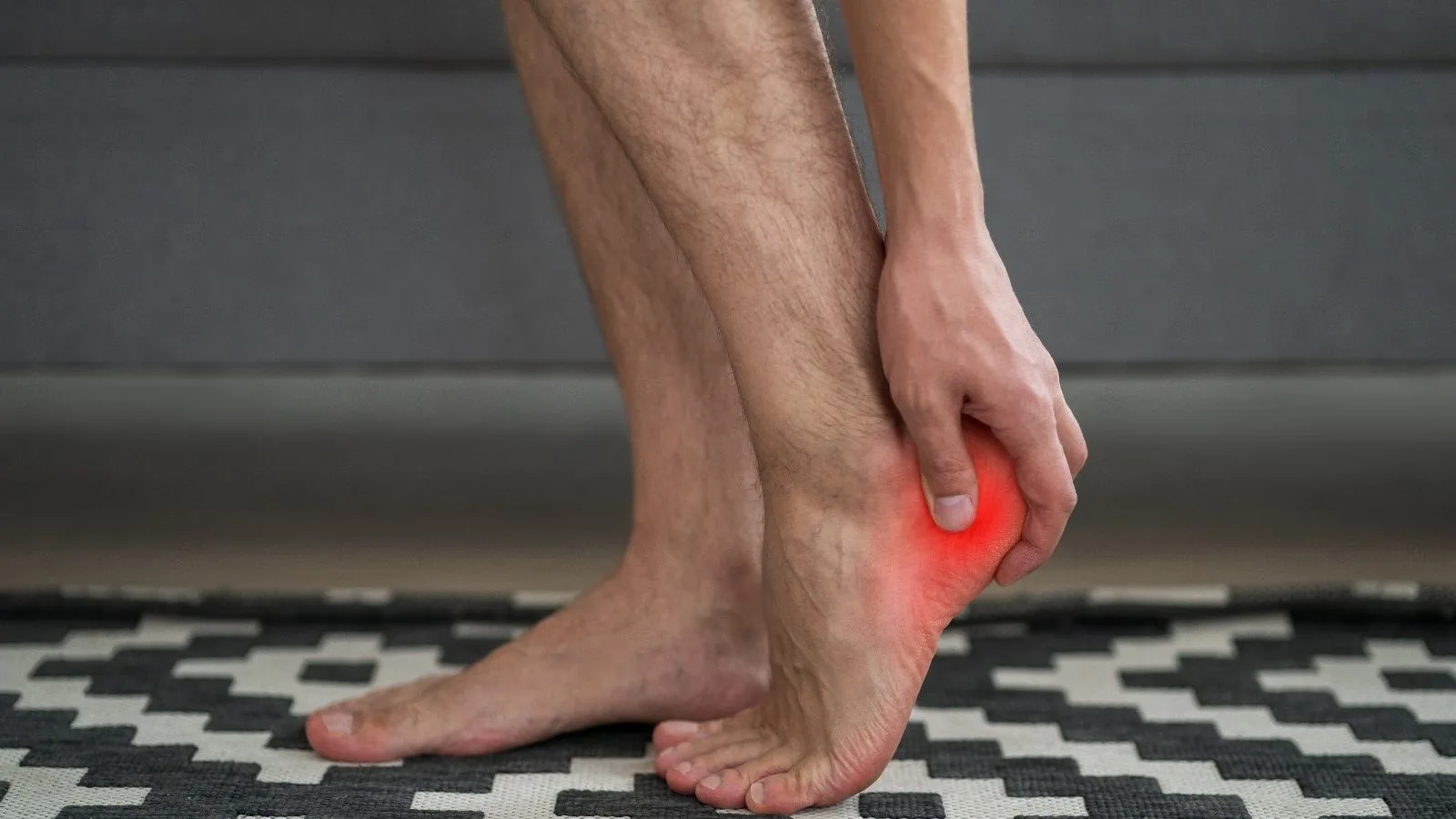
The symptoms of vascular diseases vary depending on the type of vessel affected and the stage of the disease. Pain, swelling in the legs, changes in skin color, numbness, and cramp-like pain that occurs while walking are common clinical findings. In advanced stages, signs of circulatory disorders such as delayed wound healing and a sensation of coldness may appear.
Among the causes of vascular diseases, plaque accumulation in the vessel wall, genetic predisposition, and lifestyle-related risk factors play an important role. Smoking, high cholesterol levels, diabetes, hypertension, and a sedentary lifestyle accelerate the deterioration of vascular structure. These factors can lead to narrowing of the vessels and reduced blood flow over time.
Treatment methods for vascular diseases include medical and interventional approaches planned according to the type and severity of the disease. Drug therapies, lifestyle changes, and regular exercise support circulation. In advanced cases, procedures such as angioplasty, stent placement, or surgical bypass aim to restore vessel patency.
| Things You Should Know | Information |
| Disease Group | Vascular Diseases (Vascular Disorders) |
| Subtypes | Atherosclerosis, Deep Vein Thrombosis (DVT), Varicose Veins, Peripheral Artery Disease (PAD), Aneurysm, Raynaud’s Disease, Venous Insufficiency |
| Symptoms | Pain or cramping in the legs (especially while walking), numbness, sensation of coldness, bruising, skin discoloration, swelling, varicose veins, open wounds (especially around the ankles), chest pain (in coronary vascular diseases), shortness of breath |
| Common Causes | Atherosclerosis (arterial stiffness), hypertension, diabetes, smoking, high cholesterol, obesity, genetic predisposition, sedentary lifestyle, prolonged sitting or standing |
| Risk Factors | Advanced age, family history, smoking and alcohol use, poor nutrition, insufficient physical activity, stress, hormonal changes (especially in women), certain chronic diseases (e.g., lupus, rheumatoid arthritis) |
| Diagnostic Methods | Doppler ultrasound, CT angiography, MR angiography, conventional angiography, blood tests, ankle-brachial index (ABI), ECG (in suspected coronary artery disease) |
| Treatment Methods | Lifestyle changes: regular exercise, healthy nutrition, quitting smoking; Drug therapy: blood thinners, antihypertensives, cholesterol-lowering drugs; Interventional treatment: angioplasty, stent placement; Surgical treatment: bypass surgery, vascular repairs, varicose vein surgery; Supportive treatments: use of compression stockings, physical therapy |
| Complications | Heart attack, stroke, tissue death in the legs, pulmonary embolism, organ failure, need for amputation |
| Prevention Methods | Healthy nutrition, regular exercise, quitting smoking, blood pressure and cholesterol control, diabetes management, regular medical check-ups |
Why Do Vascular Diseases Occur?
The deterioration that occurs in the vascular pathways over time is not a sudden condition; it is usually the result of a silent process that lasts for years. The most fundamental cause of diseases seen in arteries is the condition we call atherosclerosis in medicine. Cholesterol, calcium, and various cellular waste circulating in the blood begin to adhere to the inner surface of the vessel over the years. This adhesion process leads to the formation of hard, calcified structures called “plaque” in the vessel wall. This condition, resembling the narrowing of a water pipe due to limescale accumulation over time, prevents blood from passing through that area easily. The more the vessel narrows, the more the tissues that need to receive blood are deprived of oxygen and nutrients.
The main cause underlying problems in veins is the malfunction in the operating principle of the system. Veins are responsible for pushing deoxygenated blood, especially from our legs, back to the heart against gravity. While performing this challenging task, they rely on small one-way valves that prevent blood from flowing backward. However, over time, due to increased pressure or weakening of the vessel wall, these valves lose their elasticity and become unable to close properly. As the valves lose their function, blood flows backward instead of progressing toward the heart and begins to pool in the lower parts of the legs. This pooling causes the vessel to swell like a balloon under internal pressure, become twisted, and structurally deteriorate completely.
What Are the Risk Factors for Vascular Diseases?
There are many environmental and genetic factors that threaten the health of the vascular network in our body and prepare the ground for the occlusions or dilations we mentioned. While some of these factors are processes that cannot be intervened in, such as aging, the majority are directly related to lifestyle habits.
The main factors that negatively affect vascular health are listed below.
- Smoking
- Diabetes
- High blood pressure
- High cholesterol
- Advanced age
- Sedentary lifestyle
- Obesity
- Genetic predisposition
Among the items in this list, tobacco use in particular is one of the largest and most destructive triggers of vascular diseases. When toxic substances in cigarette smoke mix with the blood, they directly damage the smooth layer that lines the inner surface of the vessel and allows blood to flow easily, like a non-stick pan. When this layer is damaged, it becomes much easier for cholesterol and calcium to adhere to the vessel wall. Diabetes, on the other hand, causes damage at the cellular level by disrupting the protein and fat structures in the vessel wall over time due to persistently high blood sugar levels. While high blood pressure destroys elasticity by constantly applying intense pressure to the vessel wall from within, a sedentary lifestyle and obesity also accelerate the disease process by causing the circulatory system to become sluggish and increasing the burden on the leg veins.
With Which Symptoms Do Vascular Diseases Manifest?
The symptoms caused by vascular problems in the body vary greatly depending on whether the issue is in the arteries or veins and which organ is affected. In arterial occlusions, the main problem is cellular starvation and lack of oxygen because clean blood cannot reach the tissues. When a narrowing occurs in the leg arteries, the muscles cannot find the energy they normally need. Therefore, when a person starts walking, they feel cramp-like, compressive, and very severe pain in their calves. This pain forces the person to stop and rest. When resting, the pain subsides as the oxygen demand of the muscles decreases, but when walking resumes, the pain reappears at the same distance. In more advanced stages of the disease, a constant icy feeling in the feet, paleness or bruising of the skin, and wounds that do not heal for months even after minor trauma may occur.
In venous diseases, since the problem lies in the return of blood, symptoms are more related to pressure and fluid accumulation. Patients usually experience an increasing feeling of heaviness, fatigue, and swelling in the legs as the day progresses, especially when standing for long periods.
General complaints encountered in arterial and venous diseases are as follows.
- Calf pain
- Coldness in the feet
- Non-healing wounds
- Swelling in the legs
- Color changes
- Prominent veins
- Night cramps
Narrowing in critical vessels leading to the brain, such as the carotid artery, can present with very different and subtle symptoms. Sometimes temporary vision loss, slurred speech, or sudden loss of strength in the arm or leg may occur. These types of symptoms are very important warning signs that the brain has temporarily been deprived of oxygen or that a small fragment broken off from plaque in the vessel has reached the brain.
What Role Does Interventional Radiology Play in the Treatment of Vascular Diseases?
One of the greatest revolutions in the medical world over the past twenty years has been the development of minimally invasive methods that save patients from major surgical operations. Interventional radiology lies at the heart of this transformation. In the past, when vascular disease treatment was mentioned, the first things that came to mind were long incisions in the skin, complete removal of the vessel from the body, or replacing it with another vessel (bypass). These open surgeries meant long anesthesia times, days of hospitalization, and weeks of painful recovery for patients.
At the present point, interventional radiology uses advanced medical imaging technologies as guiding maps to look inside the body as if with a small camera system. In this way, without the need for large surgical incisions, the vascular system is accessed through a point the size of a needle hole, usually from the groin, wrist, or neck. Using the vessel’s own lumen as a pathway, the diseased area is reached directly with catheters and micro-devices thinner than a millimeter. Blocked vessels are opened from within these small tunnels, vessels at risk of rupture due to dilation are reinforced from the inside, or bleeding areas are precisely sealed. Because this modern approach does not damage tissues, it almost eliminates the risk of infection and allows the patient to return to normal life at an extraordinary speed after treatment.
Which Imaging Methods Are Used in the Diagnosis of Vascular Diseases?
The most important condition for successful and lasting treatment is identifying the source of the problem. Mapping the vascular system is the most critical stage of treatment planning. The diagnostic methods used for this purpose reveal not only the presence of the disease but also the degree of narrowing or dilation, its relationship with surrounding tissues, and the dynamics of blood flow in detail.
Color Doppler ultrasonography is one of the first-line and most reliable methods that does not involve radiation and works with sound waves in vascular health assessments. It reflects the speed, direction of blood flow inside the vessel, and plaque structures in the vessel wall instantly and in real time. It is the most valuable tool especially in varicose vein problems in the legs to determine with millimetric precision which valve is failing and allowing blood to flow backward.
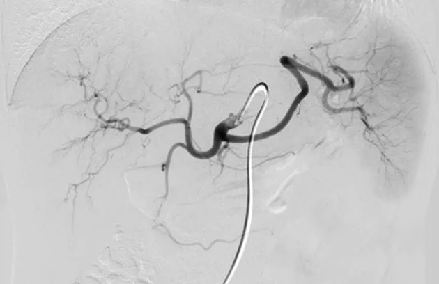
Computed tomography angiography is a technology that has revolutionized the evaluation of large vessels. With a special contrast fluid administered through a vein in the arm, a three-dimensional, high-resolution virtual map of the entire vascular network of the body is created within seconds. In cases such as vascular dilations in the abdominal region or occlusions in leg vessels, these three-dimensional images provide perfect guidance for millimetric measurements of the artificial vessel or stent to be placed. Magnetic resonance angiography performs a similar mapping using a magnetic field without radiation. Digital subtraction angiography (DSA) is the gold standard where both diagnosis and treatment can be performed simultaneously. In this method, after entering the vessel directly with a thin catheter and injecting contrast fluid, images are taken where bone and organ images outside the vessel are digitally removed, leaving only the clearest image of the vascular network, allowing even the smallest capillary problems to be detected.
How Are Leg Vessel Occlusions Treated Without Surgery?
Peripheral artery disease, commonly known as leg vessel occlusion, was previously a serious condition that could only be treated with open bypass surgery. In these surgeries, a vessel taken from another part of the body or an artificial vessel was sewn before and after the blocked area to create a new pathway for blood. Today, thanks to interventional methods, procedures to open the blocked vessel from within using its original structure have become standard.
Under local anesthesia, the blocked or narrowed area is reached through the artery in the groin with the help of special guidewires. In the first stage, balloon angioplasty is usually applied. Extremely durable micro-balloons advanced into the vessel in a deflated state are inflated at the exact center of the narrowing with externally applied pressure. The inflated balloon presses the calcified plaque narrowing the vessel against the vessel wall, reopening the tunnel required for blood flow. When the balloon is deflated, it is immediately observed that blood reaches the tissues again. However, in some cases, because the crushed plaque structure is not elastic, the vessel may collapse again after the balloon is removed. To prevent this, stents with a thin wire mesh structure are placed in the opened area. The stent acts as a permanent scaffold inside the vessel, ensuring that it remains open. In advanced cases where plaques have hardened like stone and calcified, the balloon alone is not sufficient; with special shaving devices called atherectomy, these hard plaques inside the vessel are broken down or scraped like a drill and removed from the body.
How Do Carotid Artery Diseases Trigger Stroke Risk and What Is Their Treatment?
The carotid arteries, located on both sides of our neck and carrying life-sustaining blood to the brain, are among the most common targets of atherosclerosis. Narrowing in these vessels not only reduces the amount of blood going to the brain but also carries a much more insidious and deadly danger. The calcified plaques accumulated on the vessel wall may crack over time. Microscopic fragments of calcium or clots that break off from these cracks are carried directly to the brain vessels with the blood flow. These particles, which block a capillary vessel in the brain, are one of the most important causes of stroke.
When the narrowing in the carotid artery reaches a critical level, intervention is absolutely necessary. While open surgeries involving incisions in the neck to clean the vessel were performed in the past, today safe treatment is provided through carotid artery stenting performed via the groin. The most vital part of this procedure is preventing fragments that may break off from the plaque during intervention from traveling to the brain. For this purpose, very fine “embolism protection filters” in the form of an umbrella or basket are placed beyond the narrowing. After the filter is opened, the narrowed area is expanded with a balloon and a stent is placed to trap the calcified plaque against the vessel wall. If any fragment breaks off during the procedure, it is caught in this filter before reaching the brain. After the procedure is successfully completed, the filter is collected together with these trapped hazardous particles and removed from the body.
What Is an Aortic Aneurysm and What Are the Repair Methods Without Incision?
The aorta, the largest and most important artery in our body, is like the main water supply system that carries clean blood from the heart to all our organs. This thick vessel wall, which directly withstands the enormous pressure generated with each heartbeat, can weaken over time due to aging, smoking, or genetic reasons. Especially in the abdominal section of the aorta, the weakening of the vessel wall leads to balloon-like expansion beyond its normal diameter, called an abdominal aortic aneurysm. This condition is like a ticking time bomb; when the aneurysm reaches a certain size, the vessel wall may rupture, and this internal bleeding condition often poses a life-threatening risk.
In the past, treatment of this condition involved a very large and risky open surgical operation in which the patient’s abdomen was opened from end to end, the diseased portion of the aorta was removed, and an artificial vessel was sewn in its place. Today, this life-threatening problem is resolved incision-free through the arteries in the groin using a method called “Endovascular Aortic Aneurysm Repair.” The system works as follows: With the help of catheters advanced from the groin, a stent graft (artificial vessel) consisting of a strong metal scaffold covered with a special fabric is placed inside the aorta. This artificial vessel tightly attaches to the healthy vessel wall just before and after the ballooned section. Blood flow no longer touches the old weakened vessel wall but passes entirely through this new and strong tube. Thus, the pressure on the ballooned outer area is instantly eliminated, and the risk of aneurysm rupture is permanently removed.
What Are the Current Methods in the Treatment of Varicose Veins and Venous Insufficiency?
Varicose veins, the most common form of venous system diseases, are often perceived as only a cosmetic problem, but in fact represent a serious circulatory disorder. The deterioration of the valves within the main veins responsible for carrying deoxygenated blood upward in the legs causes blood to pool below and the vessels to dilate. In the era of traditional open surgery, varicose vein treatment was based on finding this enlarged problematic vessel through incisions made from the groin and ankle and completely removing it from the leg with the help of a wire. This highly traumatic method has now been replaced by modern technologies based on the principle of closing the vessel from the inside rather than removing it.
The main non-surgical treatment options applied are listed below.
- Laser ablation
- Radiofrequency ablation
- Medical adhesive application
- Mechanochemical ablation
- Sclerotherapy
In these methods, under the guidance of an ultrasound device, the problematic vessel is accessed through a tiny entry point that does not require sutures. In laser or radiofrequency ablation methods, the vessel wall is shrunk by heat energy delivered from the tip of a thin fiber optic cable placed inside the vessel, causing it to close and become inactive. In the medical adhesive method, without using any heat, a very strong tissue glue compatible with human tissue is injected into the vessel, sealing it permanently within seconds. The function of this closed diseased vessel is immediately taken over by the numerous healthy collateral veins in the leg. For fine superficial veins visible on the surface, a drying agent is injected into the vessel with fine needles to eliminate these superficial problems.
How Are Vascular Diseases Called Deep Vein Thrombosis Treated?
Deep vein thrombosis (DVT) is a condition in which the main veins running deep between the muscles in our legs suddenly become completely blocked by a large blood clot. Since the upward flow of blood is abruptly cut off, the leg quickly swells, becomes tense, discolored, and severe pain occurs. The most frightening aspect of this condition is not the swelling of the leg, but the risk that a piece of this fresh clot may break off and travel through the bloodstream to the lung vessels, causing a blockage and stopping vital functions.
In classical treatment, patients are only given blood-thinning medications. However, blood thinners only prevent the formation of new clots; they do not dissolve the existing clot. It may take months for the body to dissolve this hard clot on its own, and during this process, the vessel valves are permanently damaged. Interventional radiologists, however, take a much more active approach to this condition. Under ultrasound guidance, a catheter is placed directly into the center of the clot via the back of the knee or the groin. With special devices, powerful drugs that immediately dissolve the clot are sprayed into it, and the clot is also mechanically broken down with rotating heads and vacuumed out of the body like a vacuum cleaner. In this way, the vessel is completely opened on the same day, the swelling of the leg subsides, and the patient is protected from future permanent circulatory disorders.
How Do Embolization Methods Treat Diseases Such as Uterine Fibroids and Varicocele?
The approach of interventional radiology to vascular diseases is not limited to opening blocked vessels. Sometimes, treatment is achieved with the opposite logic, by deliberately blocking the vessels that feed a diseased tissue or organ from within. This procedure is called embolization. Tumors, fibroids, or structurally abnormal vascular clusters require intense blood flow to grow and survive. Embolization is a highly effective method that cuts off the blood supply of these diseased structures, forcing them to shrink or regress.
Uterine fibroids, which are very common in women and cause heavy bleeding and severe pelvic pain, often led to surgeries involving complete removal of the uterus in the past. Today, with the uterine fibroid embolization procedure, the artery supplying the uterus is reached via the groin. Microscopic embolic particles are injected only into the capillary vessels feeding the fibroid mass. Fibroids that lose their blood supply shrink over months, the patient’s bleeding complaints disappear, and most importantly, the uterus remains in the body. Varicocele, a condition seen in men characterized by the dilation of veins in the scrotum, is treated with a similar approach. This abnormal reverse blood flow, which causes infertility and pain, is stopped by entering through the groin or neck vein and placing tiny platinum coils into the problematic vessel. Since there is no surgical incision, patients are discharged the same day and return to their normal lives.
What Is Done in the Processes of Diabetic Foot and Kidney Vascular Diseases?
Vascular problems can cause damage not only in specific regions of the body but also in our most sensitive organs and extremities. One of the most significant complications of diabetes in later years is diabetic foot wounds. High blood sugar eventually blocks the capillary vessels below the knee and around the ankle, preventing proper tissue nourishment. Even a small cut on the foot cannot heal due to lack of oxygen and can turn into progressively enlarging gangrenous wounds. These patients often face the risk of amputation of the foot or leg. Interventional radiologists access these below-the-knee vessels, which are too thin for standard devices, using special microcatheters and expand these millimetric narrowings with balloons. Restoring blood flow to the toes is the most critical step for wound healing and saving the limb.
The health of our kidneys is also directly dependent on the renal arteries that supply them. When narrowing occurs in these vessels, the kidney begins to secrete a hormone that constantly raises blood pressure because it cannot receive enough blood. Severe hypertension resistant to medication, especially in young individuals, is often due to these vascular narrowings. When these vessels are opened with a balloon or stent, blood pressure stabilizes and the progression toward kidney failure is halted. Similarly, in patients undergoing dialysis due to kidney failure, when the artificial vascular access (fistula) created in the arm becomes blocked by a clot, these vessels are cleared to prolong the life of the fistula and ensure uninterrupted continuation of dialysis treatment.
What Are the Preparation Before Interventional Procedures and Post-Procedure Recovery Methods?
Although interventional radiological methods do not physically strain the patient, a very meticulous clinical preparation and follow-up protocol is applied to ensure the safety of the process. Patients are asked to fast for a certain period before the operation. Detailed blood tests performed beforehand evaluate the kidneys’ ability to filter the contrast material used during the procedure and the blood clotting parameters. If necessary, the doses of blood thinners or diabetes medications used by the patient are adjusted according to the day of the procedure. In the majority of interventions, general anesthesia is not applied; only the small area where the entry will be made is numbed, and patients remain awake and able to communicate during the procedure.
Some key points that patients should pay attention to after the procedure are listed below.
- Bed rest
- Adequate fluid intake
- Avoiding heavy lifting
- Regular use of medications
After the needle used to enter through the groin or arm is removed, pressure is applied for a short time to stop bleeding, and sometimes a small plug is placed. There is no need for sutures. Patients are usually kept under observation for 4 to 6 hours after the entry site is controlled and are discharged on the same day. Fluid intake should be increased to protect the kidneys after the procedure. Regular use of prescribed medications, especially blood thinners, is critical for the long-term success of the treatment.
What Are the Advantages of Interventional Radiology Compared to Traditional Surgery?
This major transformation in modern medicine’s approach to vascular diseases has placed patient comfort and safety directly at the center of treatment. The advantages offered by interventional methods compared to open surgical operations are very clear. The greatest benefit is the avoidance of large incisions that would cause trauma to the body. Since tissue integrity is preserved, severe postoperative pain, weeks of wound care, and the risk of dangerous infections at the incision site are almost completely eliminated.
The use of local anesthesia instead of general anesthesia minimizes the burden on vital organs such as the heart and lungs. This is especially beneficial for elderly patients or those with advanced heart failure or respiratory problems, who are considered high-risk and unsuitable for open surgery, offering them a new chance at treatment. The reduction of hospital stay from weeks to hours not only provides great psychological relief but also allows patients to return to their daily life, work, and social activities as early as the next day. Preserving the natural structure of the vascular system and solving the problem from within with minimal intervention represents the most advanced stage reached in vascular medicine.
Frequently Asked Questions
What Are Vascular Diseases and how do they affect the circulatory system?
Vascular diseases are conditions that affect arteries, veins, and lymphatic vessels. They can disrupt normal blood flow, reduce oxygen delivery to tissues, and increase the risk of serious cardiovascular complications.
What symptoms may indicate the presence of vascular diseases?
Common symptoms include leg pain, swelling, numbness, skin discoloration, cold extremities, non-healing wounds, varicose veins, and fatigue. Symptoms vary depending on the location and severity of the vascular condition.
What causes vascular diseases to develop over time?
Vascular diseases can result from atherosclerosis, aging, smoking, high blood pressure, diabetes, obesity, genetic factors, inflammation, and other conditions that damage blood vessels or impair circulation.
Who is at greater risk of developing vascular diseases?
Individuals with diabetes, hypertension, high cholesterol, obesity, a family history of cardiovascular disease, or a history of smoking face a higher risk of developing vascular disorders and related complications.
Can vascular diseases increase the risk of stroke or heart attack?
Yes, certain vascular diseases can reduce or block blood flow to critical organs. This may increase the likelihood of stroke, heart attack, tissue damage, or other potentially life-threatening medical emergencies.
How are vascular diseases diagnosed by specialists?
Diagnosis may involve physical examination, Doppler ultrasound, CT angiography, MR angiography, blood tests, or catheter-based imaging studies to evaluate blood flow and identify vessel abnormalities.
What treatment methods are available for vascular diseases?
Treatment depends on the specific condition and may include lifestyle modifications, medications, minimally invasive procedures, endovascular interventions, or surgery to restore and maintain healthy circulation.
Can vascular diseases be treated with minimally invasive procedures?
Many vascular conditions can be managed through minimally invasive techniques such as angioplasty, stenting, embolization, or catheter-based therapies, which often provide shorter recovery times than traditional surgery.
How can lifestyle changes help prevent vascular diseases?
Regular exercise, a balanced diet, smoking cessation, weight management, and proper control of blood pressure, cholesterol, and diabetes can significantly reduce the risk of vascular disease progression.
Can vascular diseases lead to long-term complications if untreated?
Untreated vascular diseases may result in chronic pain, limb ischemia, ulcers, organ damage, stroke, heart attack, or permanent disability. Early diagnosis and treatment are important for improving outcomes.

Prof. Dr. Özgür Kılıçkesmez is one of Türkiye’s leading interventional radiology and neuroradiology specialists, focusing on minimally invasive endovascular treatments. His primary areas of expertise include aneurysm and stroke treatments, embolization procedures, non-surgical treatments of thyroid and parathyroid diseases, interventional oncology, and peripheral vascular interventions. He currently serves as the Head of the Interventional Radiology and Neuroradiology Department at Istanbul Memorial Göztepe Hospital.
Graduating from Istanbul University Cerrahpaşa Faculty of Medicine in 1997, Prof. Dr. Kılıçkesmez received advanced training in interventional radiology and oncology as a Clinical Fellow at London Guy’s & St Thomas’ and King’s College Hospital between 2012 and 2013. He earned the title of Professor in 2020 and, in the same year, became the Founding Interventional Radiologist at SBÜ Çam and Sakura City Hospital. He also holds the EBIR (European Board of Interventional Radiology) certification.
Prof. Dr. Kılıçkesmez has 94 international and 40 national publications and over 2500 citations. He serves as a reviewer for prestigious journals such as the American Journal of Roentgenology (AJR) and Diagnostic & Interventional Radiology. With his clinical and academic expertise, he is a nationally and internationally recognized authority in stroke treatment, brain aneurysm, embolization, tumor ablation, and minimally invasive treatments of thyroid/parathyroid diseases.





Vaka Örnekleri