


A lung biopsy is a procedure in which a tissue sample is taken from the lung to diagnose infections, inflammation, or cancer. It provides vital information when imaging alone is insufficient.
Different methods are available, including needle biopsy, bronchoscopy, and surgical biopsy. The choice depends on the location and size of the lesion, as well as the patient’s overall health.
Complications such as bleeding or pneumothorax may occur but are minimized with careful technique and imaging guidance. Most patients recover quickly after the procedure.
Lung biopsy is essential for accurate diagnosis and treatment planning. It helps physicians identify the exact cause of lung abnormalities and select the most appropriate therapy.
What Is a Lung Biopsy?
To provide detailed information about what a lung biopsy is, this procedure is a medical method used particularly in the diagnosis of lung diseases and lung cancer. It is performed to accurately diagnose abnormal conditions detected in the lungs. Lung biopsy can be performed by doctors using various techniques. During the procedure, a small sample is taken from the lung tissue and examined under a microscope. This allows doctors to determine the type and spread of the disease.
Biopsy Methods:
- Bronchoscopy: Biopsy is performed by accessing the lungs through the airways.
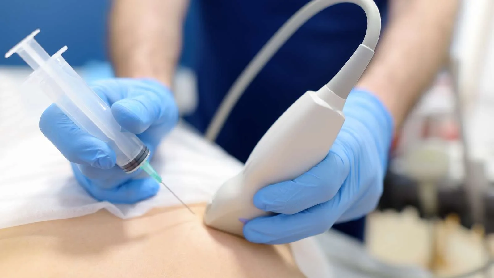
- Percutaneous biopsy: Biopsy is performed through the skin using a needle and is generally guided by computed tomography.
What Is the Purpose of a Lung Biopsy?
Lung biopsy is an effective method for the early diagnosis of diseases. It is especially used for identifying serious health problems such as lung cancer. Doctors utilize various biopsy techniques when evaluating a patient’s condition. These techniques include closed and open methods. Each is ideal for certain situations and conditions.
- Closed methods are less invasive and are generally performed under local anesthesia. Needle biopsy and transbronchial biopsy fall into this category. During needle biopsy, tissue samples are taken from the suspicious area. Transbronchial biopsy is performed through a tube inserted via the trachea.
- Open methods require a more comprehensive intervention. Procedures such as thoracoscopic biopsy are performed under general anesthesia, and an endoscopic device is inserted into the chest cavity.
Why Is a Lung Biopsy Performed?
Lung biopsy is performed to diagnose various health issues. Primarily, it is necessary to confirm abnormalities detected by imaging techniques such as tomography and chest X-ray. It also plays a critical role in identifying serious diseases such as lung infections and cancer. The causes of abnormal fluid accumulation in the lungs are also examined with this method. Biopsy provides important information when distinguishing whether suspicious tissues are benign or malignant.
- To evaluate abnormalities seen on tomography and chest X-ray.
- To diagnose lung infections, cancer, and other disorders.
- To examine unexplained fluid accumulation in the lung.
- To determine whether suspicious tissue in the lung is benign or malignant.
- To assess the spread of malignant tumors.
- To classify the existing mass.
- To gather information about the patient’s health status, the location of the lesion, and the type of problem through biopsy procedures.
What to Do Before a Lung Biopsy
Pre-procedure preparations for lung biopsy are important for patient safety and the success of the procedure. The doctor provides comprehensive information about the procedure and answers all of the patient’s questions. First, the patient must understand the procedure and sign the consent form. Reviewing the patient’s medical records and performing a general health check are essential. The patient should pay attention to the following points:
- Fasting for at least 8 hours,
- Fluid intake may be allowed in the morning if local anesthesia will be used.
The patient is also expected to provide information about any allergies. The following should be reported to the doctor:
- Any allergies to local or general anesthesia,
- Iodine or latex allergy,
- All medications and herbal supplements being used.
Patients at risk of bleeding or those taking blood thinners or aspirin should inform their doctor. Lastly, if the patient is pregnant or suspects pregnancy, this should be communicated to the doctor.
What Is the Process After a Lung Biopsy?
The post-lung biopsy process varies depending on the type of anesthesia and biopsy method used. After a biopsy performed under general anesthesia, the patient is closely monitored in the recovery room. Once a stable health condition is achieved, the patient can be discharged. A similar process is followed if local anesthesia or sedation is used; the patient’s condition is monitored until vital signs such as blood pressure, pulse, and respiration become stable.
During the recovery period after a biopsy, the following symptoms should be watched for:
- Shortness of breath
- Bloody cough
- Chest pain
- Fever and chills
- Redness or swelling at the biopsy site
If any of these symptoms are observed, the patient should contact a doctor immediately. Especially after bronchoscopic methods, patients may experience throat discomfort and difficulty swallowing, which may last for several days and is considered normal. Mild cough is also a normal reaction after transbronchial biopsy.
How Long Does a Lung Biopsy Take?
The duration of a lung biopsy generally varies from patient to patient. The procedure time:
- Is usually completed in 15 minutes to 1 hour on average.
During this time, doctors take the necessary sample from the lung tissue. After the procedure, the patient is usually observed in the hospital for a few hours. During the observation period, the patient is closely monitored for possible complications.
How Is a Lung Biopsy Performed?
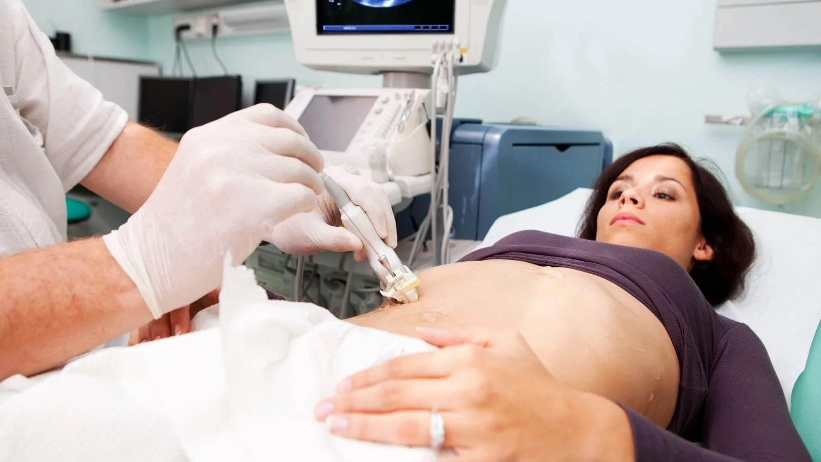
Lung biopsy is a method carefully performed by doctors. The procedure plays a significant role in assessing the patient’s health and making a diagnosis. First, the patient is evaluated by X-ray and CT scan to accurately identify the area for biopsy. After the relevant area is marked, preparation for the procedure begins:
- Jewelry or metal objects are removed.
- The patient’s clothes are changed and a hospital gown is worn.
- The area to be biopsied is cleaned with an antiseptic solution.
Local anesthesia is administered while the patient is in a comfortable position. After anesthesia, the patient is expected to remain still and control their breathing. During this time, the doctor marks the biopsy area and accesses the lung tissue with a special biopsy needle. A mild pressure sensation during the procedure is normal.
After the biopsy sample is taken, the needle is removed, and pressure is applied to the area to stop any potential bleeding. Then, a sterile bandage is applied. The tissue sample taken is sent to the pathology laboratory for further examination.
How Many Days Does It Take to Get Lung Biopsy Results?
The time it takes to obtain lung biopsy results may vary depending on several factors. The tissue sample obtained after the procedure is immediately sent to the laboratory. The workload in the laboratory and the complexity of the tissue sample are the main factors affecting the time needed for results. The nature of the tissue and the type and severity of the disease being examined may prolong the process.
- The fixation process of the tissue sample: This may sometimes take several days.
- Type of tissue: Examining hard tissues requires more time.
- Complexity of the disease: Nonspecific findings require additional testing.
Generally, biopsy results are available in 2 to 7 days. However, some complex cases may take 10 to 15 days. In any case, the examination of the tissue and the evaluation of the results are carried out with care. Pathologists perform detailed studies on the tissue and may consult other experts if necessary.
Are There Any Risks of Lung Biopsy?
The risks that may be encountered during a lung biopsy are important for every patient. Procedures such as surgical biopsy generally require general anesthesia and this brings certain risks. Complications that may be encountered during and after the procedure include:
- Blood loss and clot formation
- Infection
- Pain, tenderness, and discomfort
- Pneumonia
Less invasive methods such as needle and transbronchial biopsy are performed under local anesthesia. The following complications may occur during these procedures:
- Pneumothorax, i.e., collapsed lung
- Lung bleeding
- Infection
X-ray and CT scan devices used during lung biopsy emit a small amount of radiation. This poses a risk of radiation exposure; however, this risk is quite low. As with any surgical procedure, lung biopsy carries certain risks.
What Are the Advantages of Lung Biopsy?
Among lung biopsy methods, fine-needle biopsy stands out. This method provides many advantages and prioritizes patient comfort. The fine needles used during the procedure cause minimal tissue damage. Therefore, the patient can return to daily life shortly after the procedure. Compared to surgical intervention, fine-needle biopsy is less invasive. For patients, this means less trauma and a faster recovery process.
- Fast and Easy: The procedure is generally completed quickly, and results are obtained in a short time.
- Reliable: Analysis of the samples taken helps determine whether the tissue is benign or malignant.
- Tissue Preservation: This procedure, which is performed without the need for surgical intervention, ensures less harm to the patient.
- Diagnostic Value: The obtained cell samples are effective in identifying the type of tumor.
Patient Reviews of Lung Biopsy
For Prof. Dr. Özgür Kılıçkesmez’s reviews, see Google Maps or Doktortakvimi.
Frequently Asked Questions

How long does a lung biopsy take?
A lung biopsy usually takes between 15 and 45 minutes. This period may vary depending on the complexity of the procedure and the patient’s condition. The patient’s health status and the technique used during the biopsy affect the duration. The meticulous work of the doctors ensures that the procedure is completed within this timeframe. Therefore, the time allocated for lung biopsy varies from patient to patient depending on these factors.
Is lung biopsy painful?
Local anesthesia is generally applied to patients during lung biopsy. Thus, pain is reduced or completely prevented during the procedure. As a result, patients can have a comfortable experience throughout the biopsy. Local anesthesia prevents the sensation of pain, ensuring patient comfort. After the procedure, as the effect of anesthesia wears off, mild pain may occur, but this is usually manageable. Therefore, lung biopsy is considered a painless procedure when performed under anesthesia.
Is it dangerous to take a sample from the lung?
Taking a sample from the lung is generally considered a safe procedure. However, as with any interventional procedure, there are some risks. The most common complications are bleeding and pneumothorax. Especially in people with lung diseases such as emphysema, the risk of pneumothorax is higher and this can cause serious breathing difficulties. Therefore, the procedure requires specific preparations and it is important for the patient to follow the doctor’s recommendations.
Does biopsy spread cancer?
Biopsy is a method used to diagnose cancer. During this procedure, a small sample is taken from the tissue. However, this procedure does not cause cancer to spread in the body. Both surgical and needle biopsies are safe. The samples taken with these methods do not cause the disease to spread to other areas. Therefore, patients should not worry that the biopsy procedure will worsen the cancer. The only exception is primary cancer of the liver, where spread along the needle tract is rare, but methods are used to prevent this (coaxial technique), and it responds well to treatment.
What does a negative lung biopsy result mean?
A negative lung biopsy result means that no cancer cells were found in the examined tissue. Therefore, this result indicates that there is no cancer in the biopsied area. This type of result is especially important for guiding treatment in patients diagnosed with cancer. A negative biopsy result provides clearer information about the patient’s current condition and allows doctors to consider other possibilities besides cancer.
Why is a lung biopsy requested?
Lung biopsy is requested to evaluate abnormalities detected in chest X-ray or CT scan. It is also necessary to identify the cause of fluid accumulation in the lungs of unknown origin. This procedure plays an important role in diagnosing lung infections or other diseases. Biopsy is also performed to determine whether a lung mass is benign or cancerous. Finally, it helps shape treatment options by determining the stages of malignant tumors.
In which situations is a lung biopsy performed?
Lung biopsy is performed to diagnose new or growing solitary lung nodules, unexplained lung infiltrates where infection cannot be detected, undefined mediastinal masses, or to confirm or stage malignancies such as lung cancer. It is preferred when less invasive methods such as bronchoscopy are insufficient or when a tissue sample is needed for targeted therapy decisions. It is used when suspicious areas are detected in imaging studies and a definitive diagnosis is required for treatment planning.
What techniques are used during the biopsy procedure?
Different techniques are used during lung biopsy: Computed Tomography (CT)-Guided Percutaneous Transthoracic Needle Biopsy (PTNB): The needle is directed to the lesion through the chest wall using CT imaging; it offers 92.5% sensitivity and 98% specificity, with a 23% risk of pneumothorax and 8% risk of bleeding. Electromagnetic Navigation Bronchoscopy (ENB): Bronchoscopy is used to access peripheral lung lesions, with a 79.8% diagnostic rate and 5.2% pneumothorax risk. Endobronchial Ultrasound-Guided Transbronchial Needle Aspiration (EBUS-TBNA): Samples are taken from masses near the airways using bronchoscopy and ultrasound; it offers an 86% diagnostic rate, 0.61% risk of bleeding, 0.37% risk of pneumothorax, and 0.30% risk of infection. Mediastinoscopy: Provides surgical access to mediastinal lymph nodes; 92.8% diagnostic rate and 0.19% risk of hoarseness. Transbronchial Lung Cryobiopsy (TBLC): Larger tissue samples are taken, with 97.5% alveolar tissue yield; bleeding and pneumothorax risks exist. The choice of technique depends on factors such as lesion location, size, and the patient’s health status.
Is pain felt during a lung biopsy?
Patients generally feel mild discomfort during a lung biopsy. In a study of 215 people, 20% reported no pain, 67.9% reported mild pain (1-3/10), 11.2% reported moderate pain (4-6/10), and only 0.9% reported severe pain (7-10/10). Larger lesion size, wider needle-pleura angle, and longer procedure duration can increase pain levels. In another study, 20% of patients described the procedure as painful, while 29% experienced short-term shortness of breath after the procedure. Overall, although most patients reported minimal or mild pain, the experience may vary from person to person.
What should patients pay attention to after the procedure?
After lung biopsy, patients should be aware of pneumothorax symptoms, which can be seen in 20.5% of cases, and remember that a chest tube may be needed in 3.1% of cases. Hemoptysis occurs in 5.3% of cases. Mild pain in the biopsy area is normal, but infection symptoms (redness, swelling, discharge) should be monitored, and a doctor should be consulted if necessary. Heavy lifting and strenuous activities should be avoided for the first 3 days. Showering is generally allowed after 24 hours, but submerging the biopsy area in water should be avoided for 3 days. Since pressure changes can affect healing, flying and diving should be avoided for a week. If sudden chest pain, shortness of breath, or a large amount of blood is coughed up, seek medical attention immediately.
How long does it take to get biopsy results?
The time to obtain lung biopsy results varies depending on the type of biopsy and the analyses required. Routine biopsy results are usually ready within a few days, while more complex analyses such as molecular testing may take longer. For example, next-generation sequencing (NGS) results usually take 10-12 days, but sometimes up to 2.5-3 weeks. Liquid biopsies, which analyze circulating tumor DNA, generally provide results about 10 days faster than conventional tissue biopsies. Delays may occur if additional procedures, special staining, or a second opinion are required. Therefore, while some biopsy results are available within a few days, those requiring special tests may take several weeks.
What are the absolute contraindications or reasons to postpone a lung biopsy?
The applicability of lung biopsy may be restricted or risky in some cases. Especially in patients with uncorrectable bleeding disorders (e.g., low platelet count or clotting factor deficiency), biopsy significantly increases the risk of bleeding. Uncontrolled severe cough can make needle placement difficult and increase the risk of lung tissue damage during the procedure. Severe hypoxemia (very low blood oxygen level) or unstable cardiovascular conditions are also significant risk factors. In addition, if the patient cannot maintain the required position or cooperate during the procedure, a safe biopsy cannot be performed. In such cases, doctors may consider alternative diagnostic methods or postpone the procedure until the patient’s condition is stabilized.
What are less invasive diagnostic methods that can be used as alternatives to lung biopsy?
Although lung biopsy is the gold standard for a definitive diagnosis, less invasive methods may be considered in some cases or according to patient preference. For example, sputum cytology can be used to detect cancer cells, especially in tumors close to the central airways. Liquid biopsy (genetic tests using samples from blood or other body fluids) is increasingly important for profiling the tumor and guiding treatment, especially in advanced-stage lung cancer. Advanced imaging techniques such as PET-CT can show the metabolic activity of a lesion and help differentiate benign from malignant masses. However, these methods do not always replace biopsy and may be insufficient for diagnosis; a tissue sample may still be required in such cases.
What should patients do to accelerate recovery and reduce the risk of complications after lung biopsy?
It is critical to follow doctor’s advice to accelerate recovery and minimize possible complications after lung biopsy. For the first few days after the procedure, avoid strenuous physical activities, straining, and heavy lifting. If there is a cough, mild cough suppressants may be used as recommended by the doctor, but in cases of severe or bloody cough, seek medical attention immediately. Keeping the biopsy area clean and dry reduces the risk of infection. Drinking plenty of fluids and eating a balanced diet contribute to overall recovery. If the patient smokes, quitting or at least pausing is very important for lung healing. Attend all scheduled follow-up appointments and promptly report any unexpected symptoms (fever, increasing shortness of breath, chest pain).
What alternative diagnostic methods are used if the lung biopsy result is insufficient (non-diagnostic)?
If the lung biopsy result is insufficient, meaning the tissue sample does not contain enough cells or material for diagnosis, doctors usually follow several different steps. First, the reason for the insufficient biopsy is evaluated; factors such as small lesion size, difficult location, or the needle not reaching the correct area may be involved. Depending on the situation, the biopsy procedure may be repeated, preferably using a different imaging technique (e.g., ultrasound-guided or a different bronchoscopic method) for more targeted sampling. If repeated biopsies are still inconclusive or the patient’s condition is not suitable for repeated invasive procedures, a more comprehensive surgical biopsy (e.g., video-assisted thoracoscopic surgery – VATS) may be considered. Alternatively, depending on the lesion’s characteristics and the patient’s general condition, a period of close monitoring with imaging may be chosen.
What is the risk of ‘pneumothorax’ (collapsed lung) often mentioned after lung biopsy, and how is it managed if it develops?
Pneumothorax is one of the most common complications after lung biopsy and is the partial or complete collapse of the lung due to air leaking between the lung membrane and chest wall. The risk varies according to the biopsy method and the patient’s lung structure. Symptoms include sudden chest pain, shortness of breath, rapid breathing, and cough. Most small pneumothorax cases resolve on their own and only observation is needed; rest and sometimes oxygen therapy are recommended. However, if the collapse is significant or if the patient has serious breathing problems, a thin tube (chest tube) is placed into the chest cavity to remove the accumulated air and allow the lung to re-expand. This is usually done under local anesthesia and may require the patient to stay in the hospital for a few days.
Additional Resources and Documents

Prof. Dr. Özgür Kılıçkesmez is one of Türkiye’s leading interventional radiology and neuroradiology specialists, focusing on minimally invasive endovascular treatments. His primary areas of expertise include aneurysm and stroke treatments, embolization procedures, non-surgical treatments of thyroid and parathyroid diseases, interventional oncology, and peripheral vascular interventions. He currently serves as the Head of the Interventional Radiology and Neuroradiology Department at Istanbul Memorial Göztepe Hospital.
Graduating from Istanbul University Cerrahpaşa Faculty of Medicine in 1997, Prof. Dr. Kılıçkesmez received advanced training in interventional radiology and oncology as a Clinical Fellow at London Guy’s & St Thomas’ and King’s College Hospital between 2012 and 2013. He earned the title of Professor in 2020 and, in the same year, became the Founding Interventional Radiologist at SBÜ Çam and Sakura City Hospital. He also holds the EBIR (European Board of Interventional Radiology) certification.
Prof. Dr. Kılıçkesmez has 94 international and 40 national publications and over 2500 citations. He serves as a reviewer for prestigious journals such as the American Journal of Roentgenology (AJR) and Diagnostic & Interventional Radiology. With his clinical and academic expertise, he is a nationally and internationally recognized authority in stroke treatment, brain aneurysm, embolization, tumor ablation, and minimally invasive treatments of thyroid/parathyroid diseases.




Vaka Örnekleri