


The spinal cord is a cylindrical nerve tissue that extends within the spinal canal and enables communication between the brain and the body as part of the central nervous system. It plays a fundamental role in regulating reflexes, movement, and sensory perception by transmitting motor and sensory signals from the brain to the relevant organs and tissues.
The anatomical structure of the spinal cord consists of segments extending from the cervical region to the lumbar level and the spinal nerves emerging from these segments. This structure, composed of gray and white matter, organizes motor and sensory transmission through anterior and posterior roots and ensures segmental integrity.
The functions of the spinal cord include controlling voluntary muscle movements, transmitting sensations such as pain and touch to the brain, and generating reflex responses. Thanks to the reflex arc mechanism, some responses occur rapidly without brain involvement, contributing to the protection of the organism.
Spinal cord diseases and injuries may develop due to trauma, tumors, infections, or degenerative processes and can lead to serious neurological outcomes such as paralysis, loss of sensation, or urinary control disorders. Early diagnosis and appropriate treatment are critically important in limiting functional loss.
| Things You Need to Know | Information |
| Definition of the Spinal Cord | The spinal cord is a part of the central nervous system and is a long, cylindrical nerve tissue that transmits signals between the brain and the body. It is located within the spinal canal. |
| Anatomical Structure | The spinal cord is a structure protected by the vertebral bones (vertebrae) and extends from the brainstem to the coccyx. The point where it connects to the brain is the medulla oblongata. |
| Length | In an adult human, the spinal cord is approximately 42-45 cm long. |
| Sections | It is divided into five regions: cervical (neck), thoracic (chest), lumbar (lower back), sacral (sacrum), and coccygeal (tailbone). |
| Nerves in the Spinal Cord | 31 pairs of spinal nerves emerge from the spinal cord; these nerves provide communication between the spinal cord and the body. |
| Main Function | To transmit motor commands from the brain to the body and carry sensory information from the body to the brain. |
| Reflex Center | The spinal cord is the center of some reflex movements. These reflexes occur directly at the spinal cord level, independently of the brain. |
| Transmission Function | There are ascending (afferent) and descending (efferent) nerve pathways along the spinal cord. These pathways carry information. |
| Protective Structures | The vertebral bones, cerebrospinal fluid (CSF), and spinal membranes (meninges) protect the spinal cord from external factors. |
| Diseases and Injuries | Spinal cord injuries can lead to paralysis, sensory loss, or organ dysfunction. Conditions such as multiple sclerosis, spinal cord tumors, and trauma can affect the spinal cord. |
Where Is the Spinal Cord Located in Our Body?
This vital nerve tissue, which begins at the major connection point just below our skull, descends down our back and usually ends in the upper parts of our lower back region. In an adult human, this structure, which is on average about forty-five centimeters long, resembles a delicate cord about the thickness of a finger. Protecting such a critical and delicate structure from external factors is of vital importance. Therefore, nature has placed it inside a perfect armor. This armor is the series of bones extending along our back, namely our spine. When the openings in the middle of our vertebral bones align, they form a secure canal, and our nerve network passes through this protected tunnel.
As it progresses within this protective bone structure, branches begin to extend to the right and left from various points along the cord. These branches are nerve roots that reach the farthest points of our body. Everything, from the sense of touch at our fingertips to the muscles that move our toes, is connected to the central system through these branches.
What Sections Does Our Spinal System Consist Of?
Our spine is not a single solid bone but consists of a total of thirty-three separate vertebrae that provide mobility and protect the nerve tissue inside from mechanical trauma. Our main spinal regions, classified according to their position in the body, are as follows:
- Cervical region
- Thoracic region
- Lumbar region
- Sacral region
- Coccygeal region
The cervical region consists of the first seven bones that form our neck. This section, which allows our head to move in all directions, also contains very vital blood vessels leading to the brain within its small openings. Conditions we call cervical disc herniation occur due to the wear of cartilage in this region.
The thoracic region consists of twelve bones that form our back. These bones connect with our ribs to form the rib cage. They provide a strong structure that protects our internal organs, our heart, and our lungs. The spinal canal in this region is slightly narrower than in other regions, so even a minor problem here can quickly put pressure on the nerves.
The lumbar region consists of five large bones that form our lower back. This region carries almost all of our body weight. Since it is the area most exposed to physical strain such as bending, standing up, and lifting loads, it is the most common site for problems such as lumbar disc herniation and spinal canal narrowing.
The sacral and coccygeal regions form the lowest part of the spine. The five bones in the sacral region are fused together and connect with the pelvis, balancing the center of gravity of our body. The coccygeal region, or tailbone, is the final part that supports us when we sit.
What Are the Membrane Layers That Protect the Spinal Cord?
The protection provided by bones alone is not sufficient for this delicate nerve structure. On the inner surface of the bony tunnel formed by the spine, there is a special three-layered sheath system that completely surrounds the cord. These membrane layers, listed from outer to inner, are:
- Dura mater
- Arachnoid mater
- Pia mater
The dura mater is the outermost membrane layer, extremely tough, durable, and inelastic. Its purpose is to protect the delicate structure inside from friction and impacts coming from the bones. The space between the dura mater and the vertebral bones is called the epidural space. This is the safe area where we can administer medications in pain treatments just before reaching the nerve roots.
The arachnoid mater is the middle membrane located just beneath the dura mater and consists of fine connections that resemble a spider web when examined under a microscope.
The pia mater is the innermost membrane, tightly adhered to the surface of the nerve tissue and containing the capillaries that nourish it. It is the thinnest and most delicate layer.
A very special fluid circulates between the arachnoid and pia membranes. This clear fluid, called cerebrospinal fluid, suspends the entire cord within it. This fluid acts like a perfect hydraulic shock absorber system that prevents shocks generated when we walk, run, or jump from damaging nerve cells. At the same time, it is responsible for nourishing the tissue and removing waste.
What Are the Main Functions of the Spinal Cord in Our Body?
This system, which functions like a complex telephone exchange or a massive computer server, has vital functions. Its main duties are as follows:
- Sensory signal transmission
- Motor command transmission
- Reflex center control
- Autonomic system regulation
Sensory signal transmission is the basis of our physical interaction with our environment. When you hold a hot cup of coffee, feel the wind touching your skin, or feel pain when stepping on a sharp object, these data are received from the nerve endings in the relevant region. This electrical information traveling along the nerves enters from the posterior part of the spinal cord and is rapidly carried upward to the brain. Interestingly, these signals follow a crosswise path inside. In other words, the pain from a thorn pricking your left foot crosses to the right side within the cord and is transmitted to the right hemisphere of the brain.
Motor command transmission is the system that enables us to move. A desire to walk, run, or write formed in the movement planning center of our brain is sent downward as electrical signals. These commands exit from the anterior parts of the cord, reach the relevant muscle group, and initiate contraction. Any disruption or compression in these pathways can lead to loss of strength known as paralysis.
Reflex center control is the emergency braking system that protects our body from sudden dangers. The time required to withdraw your hand quickly when touching a very hot object is extremely short. If this signal first went to the brain, was processed, and then returned, your hand would already be burned. Instead, when the pain signal reaches the spinal cord, a motor command is immediately generated there without consulting the brain, and the muscles are instructed to “withdraw.” This is a completely involuntary and life-saving rapid mechanism.
How Are Spinal and Spinal Cord Diseases Diagnosed in Radiology?
In cases of any pain, numbness, or loss of strength, we must visualize the source of the problem to plan the correct treatment. When our patients apply to us, we use various high-technology devices to determine the issue. The diagnostic methods we frequently use today are:
- Magnetic Resonance Imaging
- Computed Tomography
- Digital Angiography
- Dynamic Fluoroscopy
Magnetic Resonance Imaging, or MRI, is our gold standard for showing soft tissues with tremendous clarity. With MRI, we can examine in slices how much a herniation presses on a nerve, the degree of canal narrowing, whether there is a tumor or infection inside, and even a millimetric edema within the cord. Moreover, since it does not use X-rays, it does not carry radiation risk.
Computed Tomography is an excellent tool for examining bone structures. We benefit from CT scans in compression fractures caused by osteoporosis, calcifications, and structural abnormalities in the vertebral bones. It allows us to measure the extent of damage by creating a three-dimensional map of the bones.
Digital Angiography and Dynamic Fluoroscopy devices are mostly used by interventional radiologists like hands and eyes during treatment. With special dyes administered into the vessels, we can monitor blood flow in real time and track the direction of our therapeutic needles on the screen with millimetric precision.
What Are Non-Surgical Treatments Applied in Spinal Fractures?
Compression-type collapse fractures may occur in spinal bones due to advanced age, severe osteoporosis, or trauma. These fractures cause severe back pain that can make even breathing unbearable. In the past, these patients had to remain bedridden for months with steel braces. However, today we apply very comfortable cementation techniques performed through a needle hole. These treatment methods are:
- Vertebroplasty
- Kyphoplasty
These techniques are not open surgeries. No incision is made on the skin, no scalpel is used, and no stitches are applied. Under the guidance of imaging devices, the fracture area is repaired from the inside by entering the affected bone with very fine systems. Patients can stand up and walk on the same day.
What Are the Basic Steps of the Vertebroplasty Procedure?
This procedure is performed under local anesthesia and mild sedation to relax the patient. While the patient lies face down, the exact location of the fractured bone is marked on the screen using a mobile X-ray device called fluoroscopy. A tool as thin as the tip of a needle is inserted directly into the collapsed bone from the back.
When we reach the core of the bone, we slowly inject a substance known as polymethylmethacrylate, commonly referred to as “bone cement,” while it is in liquid form using a special syringe. This fluid fills the fracture cracks inside the bone. Within just a few minutes, it hardens like concrete. During this hardening process, the slight heat generated also suppresses the pain nerves in that area. Since the fracture is frozen and stabilized from the inside, the patient’s severe mechanical bone pain disappears almost miraculously as soon as the procedure is completed.
What Are the Differences Between Kyphoplasty and Vertebroplasty?
The kyphoplasty method is a more advanced and improved version of vertebroplasty. We prefer this method especially in cases where the collapse in the bone is significant and the patient has begun to develop a hunchback.
In this procedure, instead of directly injecting cement, we place a very strong, specially designed medical balloon into the bone in a collapsed state. By slowly inflating the balloon inside the bone, we restore the crushed and collapsed bone to its original height. In this way, the posture disorder in that region of the spine is corrected.
Once the bone returns to its original shape, the balloon is deflated and removed. The cavity created by the balloon is then filled with bone cement at a much lower pressure. The greatest advantage of the procedure is that, in addition to restoring bone height, it almost eliminates the risk of cement leakage into vessels or the spinal canal.
What Are Targeted Treatments Applied in Lumbar and Cervical Disc Herniation?
In the vast majority of patients presenting with lumbar or cervical disc herniation complaints, there is no need for surgery to solve the problem. To control this inflammatory and painful condition caused by the rupture of the cushions between the vertebrae and compression of the nerves, we perform targeted drug applications directly at the source. The most effective targeted treatment methods we apply for herniation pain are:
- Epidural steroid injections
- Transforaminal epidural injections
- Facet joint injections
These procedures are not performed blindly or in a simple clinical setting. They are always performed under X-ray guidance in an angiography or fluoroscopy unit. After identifying the exact location of the problematic nerve root or spinal canal on the screen, we reach the target with a millimetric needle. To ensure the needle is in the correct place, we inject a small amount of contrast dye and confirm its spread on the screen. Then we deliver a powerful mixture of corticosteroid and local anesthetic directly to the area.
What Is the Effect of Epidural Steroid Injections on the Patient?
When a herniation ruptures or when the fluid inside the disc leaks out, the nerve root passing through that area shows a severe reaction to this foreign substance. The nerve becomes red, swollen, and edematous. The main cause of the electric shock-like sensation radiating to the leg or the unbearable pain spreading from the neck to the arm is this mechanical compression and chemical edema.
The powerful drug mixture we deliver to the affected area suppresses the inflammatory reaction like a fire extinguisher within seconds. As the edema resolves, the pressure on the nerve decreases, the swelling subsides, and the nerve begins to “breathe” comfortably again. The patient feels a significant reduction in pain even while getting up from the table. While oral muscle relaxants or painkillers may strain the stomach and kidneys and only provide a general effect, this method allows direct intervention at the very core of the inflammation.
How Does Radiofrequency Treatment Provide a Solution for Facet Joint Pain?
The small joints that connect our spinal bones at the back and allow us to move, just like our knees or elbows, are called facet joints. Over time, due to poor posture, excess weight, or aging, severe degeneration can occur in these small joints. The stiffness you feel in your lower back when you wake up in the morning or the stabbing pain you feel when leaning backward usually originates from these joints.
Radiofrequency treatment targets the small nerve branches that transmit pain from these degenerated joints. The procedure is again performed under imaging guidance. Using a very thin electrode needle, we approach the nerve fiber that carries the pain. Radio waves sent from a special radiofrequency generator create a small, controlled heat energy at the tip of the needle. This heat, reaching approximately eighty degrees Celsius, desensitizes the nerve carrying the pain at the cellular level.
Since the nerve communication pathway is interrupted, even if degeneration continues in that area, the patient no longer feels pain. Its effect usually lasts from six months to one and a half years, providing highly satisfactory and nearly long-lasting comfort.
What Are Disk Treatments with Laser and Plasma Technology?
Thanks to advances in medical engineering, we can now not only administer medication externally in spinal treatments but also reduce the volume of the herniated cartilage from the inside without creating an open wound. The next-generation technological treatments we apply in moderate herniations that have not yet ruptured are:
- Percutaneous laser disk decompression
- Radiofrequency nucleoplasty
- Ozone injection
All of these methods are performed through a single entry point under local anesthesia applied to the skin. Patients can return to their normal daily activities on the same day. In cases where the outer layer of the disc (annulus fibrosus) is intact but bulging, these procedures yield excellent results.
What Is Percutaneous Laser Disk Decompression and How Does It Work?
This method, abbreviated as PLDD in the medical field, is based on changing the mechanical dynamics of the herniation. When the pressure inside the cartilage structure (disc) between two vertebrae increases significantly, it bulges outward and narrows the nerve canal.
In this procedure, we reach the core of this cartilage disc by inserting a very thin, long needle through the patient’s lower back or neck. Through this needle, we send a microscopically thin laser fiber optic cable. When the laser energy is activated, it vaporizes a very small portion of the gelatinous tissue at the center of the disc within seconds using high heat.
The main goal here is not to completely eliminate the disc. When a small amount of fluid at the center of the disc evaporates, the internal pressure within the closed system of the disc suddenly drops. This sudden pressure drop creates a strong vacuum effect inside. Just like a deflated tire, the herniated portion that has protruded outward is pulled back inward by this vacuum force. As the external pressure disappears, the compression of the nerve by the herniation stops.
How Is Plasma Technology Used in Nucleoplasty Procedures?
In the nucleoplasty method, developed as an alternative to laser technology, the main energy source is radio waves. With specially designed, flexible-tip smart electrodes, we enter the herniated disc.
The energy delivered from the tip of this system creates a plasma field in the surrounding tissue. Unlike laser, nucleoplasty does not use very high temperatures. At relatively low temperatures of around forty to fifty degrees Celsius, it breaks the molecular bonds of tissue proteins inside the disc and converts the tissue into gas. The electrode is advanced through several different channels within the cartilage, creating small cavities inside. These cavities allow the cartilage to shrink and lose volume. Since nucleoplasty is performed in a very narrow area in cervical herniations, it is an extremely safe alternative as it prevents heat from damaging surrounding tissues.
What Are Vascular Diseases Around the Spinal Cord?
For the spinal cord, which we call medulla spinalis, to function properly, it requires a continuous flow of oxygen and nutrients. This flow is provided by a highly complex and delicate vascular network. However, due to congenital or acquired causes, abnormalities may occur in this vascular network. The most serious vascular diseases we encounter are:
- Arteriovenous malformations
- Dural arteriovenous fistulas
- Spinal cord aneurysms
- Spinal vascular occlusions
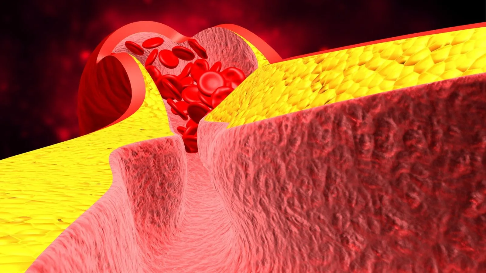
These diseases are among the most dangerous and insidious problems of the spine and nervous system. The formation of vascular tangles (malformations) or abnormal connections between arteries and veins (fistulas) completely disrupts the blood pressure in the region.
Since veins cannot carry high-pressure blood, they expand, swell, and begin to twist like a snake, exerting physical pressure on the spinal cord. Worse still, these thin-walled vessels can suddenly rupture and bleed into the spinal cord. Patients may experience gradually worsening symptoms such as weakness in the legs, urinary incontinence, or numbness over months, or they may present with sudden paralysis within seconds due to acute bleeding.
How Are Spinal Angiography and Embolization Procedures Performed?
Treating such deep and complex vascular tangles with open surgery is extremely difficult and requires opening the patient’s back entirely, with a very high risk of nerve damage. Interventional radiology has created a major revolution in medicine at this point. We can now treat vascular diseases of the spinal cord directly from within the vessel through a needle hole.
The patient is taken to the angiography unit. Under local anesthesia, a small plastic tube (sheath) of just one or two millimeters is inserted into the artery in the groin area. Through this entry point, special wires and catheters (thin, long medical tubes) are advanced upward along the main artery of the body.
By entering the fine neck and back vessels that supply blood to the spinal cord one by one, contrast dye is injected, and a detailed map of the vascular network is obtained on large screens. Once the problematic tangle or abnormal connection is identified, very special tubes called microcatheters, slightly thicker than a strand of hair, are advanced directly into the core of this diseased tangle.
When the target is reached, we permanently close the abnormal vessels using liquid embolic agents, that is, medical adhesives. This liquid substance solidifies instantly upon contact with the vessel, forming a plastic-like plug. Blood flow returns to normal, the swollen vessels shrink, and the pressure on the spinal cord is eliminated. This method, performed without opening the back or cutting bones, is today the primary treatment option for vascular spinal cord diseases.
What Is Image-Guided Biopsy in Spinal Tumors and Infections?
Masses of unknown origin, suspected tumors that may have metastasized from other organs, or persistent infection foci can sometimes be detected in the spinal bones or surrounding muscle tissues. In order for oncology and infectious disease specialists to administer the correct chemotherapy, radiotherapy, or appropriate antibiotic treatment, it is essential to establish a laboratory diagnosis of the tissue, that is, to obtain a tissue sample.
In the past, obtaining this sample required opening the relevant area under operating room conditions. Today, in interventional radiology, we perform this entirely with closed methods. The main biopsy guidance methods we use are:
- Ultrasound guidance
- Computed tomography guidance
- Fluoroscopy guidance
With the tomography device, the three-dimensional position of the lesion and the surrounding vital vessels and nerves is clarified. Thanks to millimetric measurements on the screen, the safest access angle to the target is determined. A thin, long needle is inserted through the skin, passing through safe tissues, and reaching directly into the center of the mass. Using a biopsy needle with a special nested mechanism, small cylindrical samples are taken from the mass.
In this way, the risk of damaging nerves is eliminated, and the patient can return home the same day to await the pathology result. Large surgical wounds and infection risks are avoided with this method.
What Are the Advantages of Interventional Radiology Procedures Compared to Open Surgery?
At this stage reached by modern medicine, “interventional radiology” has become a discipline that completely transforms the classical understanding of surgery and provides maximum comfort and safety to the patient. These minimally invasive procedures performed on an extremely delicate area such as the spine and spinal cord have numerous advantages. The main advantages are:
- No incision, sutures, or scalpel marks
- Prevention of long hospital stays
- Minimization of infection risk
- No damage to surrounding healthy tissues
- Elimination of the necessity for general anesthesia
Since large incisions are not made and muscle tissues are not cut and separated, there is no healing process like in traditional surgery. The patient does not experience postoperative pain when getting up from the table. Since most procedures are performed under local anesthesia and mild sedative medications, high-risk patients such as the elderly or those with heart or respiratory failure can be treated safely. Most patients can walk into the hospital and leave a few hours after the procedure, again walking and relieved of their pain. This situation also keeps the patient’s morale extremely high psychologically.
Frequently Asked Questions
What Is the Spinal Cord and where is it located in the body?
The spinal cord is a long bundle of nerve tissue that extends from the brain through the spinal canal. It serves as the primary communication pathway between the brain and the rest of the body.
What Are the Functions of the Spinal Cord in the nervous system?
The spinal cord transmits sensory information from the body to the brain and carries motor signals from the brain to muscles. It also coordinates many reflex actions without direct brain involvement.
How does the spinal cord help control movement and coordination?
The spinal cord carries nerve impulses that allow muscles to contract and perform voluntary movements. It plays a crucial role in balance, posture, coordination, and the execution of everyday physical activities.
What sensory information is transmitted through the spinal cord?
The spinal cord relays sensations such as touch, pain, temperature, pressure, and body position from peripheral nerves to the brain, allowing the body to respond appropriately to environmental stimuli.
What conditions can affect the spinal cord and its function?
Spinal cord function can be affected by trauma, herniated discs, tumors, infections, vascular disorders, inflammatory diseases, and degenerative conditions that interfere with nerve signal transmission.
What symptoms may indicate a spinal cord disorder or injury?
Symptoms may include weakness, numbness, tingling, loss of coordination, difficulty walking, chronic pain, bowel or bladder dysfunction, and, in severe cases, partial or complete paralysis.
How are spinal cord conditions diagnosed by specialists?
Diagnosis typically involves neurological examinations, MRI scans, CT scans, X-rays, electromyography, and other imaging or diagnostic tests that evaluate the structure and function of the spinal cord.
Can spinal cord injuries lead to permanent neurological deficits?
The outcome depends on the severity and location of the injury. Some patients recover partially or fully, while others may experience long-term neurological impairments that require ongoing rehabilitation and support.
What treatment options are available for spinal cord disorders?
Treatment may include medications, physical therapy, rehabilitation, minimally invasive procedures, surgery, or multidisciplinary care aimed at relieving symptoms and preserving neurological function.
How can spinal cord health be protected throughout life?
Maintaining good posture, exercising regularly, avoiding smoking, preventing injuries, managing chronic health conditions, and seeking prompt medical attention for neurological symptoms can help support spinal cord health.

Prof. Dr. Özgür Kılıçkesmez is one of Türkiye’s leading interventional radiology and neuroradiology specialists, focusing on minimally invasive endovascular treatments. His primary areas of expertise include aneurysm and stroke treatments, embolization procedures, non-surgical treatments of thyroid and parathyroid diseases, interventional oncology, and peripheral vascular interventions. He currently serves as the Head of the Interventional Radiology and Neuroradiology Department at Istanbul Memorial Göztepe Hospital.
Graduating from Istanbul University Cerrahpaşa Faculty of Medicine in 1997, Prof. Dr. Kılıçkesmez received advanced training in interventional radiology and oncology as a Clinical Fellow at London Guy’s & St Thomas’ and King’s College Hospital between 2012 and 2013. He earned the title of Professor in 2020 and, in the same year, became the Founding Interventional Radiologist at SBÜ Çam and Sakura City Hospital. He also holds the EBIR (European Board of Interventional Radiology) certification.
Prof. Dr. Kılıçkesmez has 94 international and 40 national publications and over 2500 citations. He serves as a reviewer for prestigious journals such as the American Journal of Roentgenology (AJR) and Diagnostic & Interventional Radiology. With his clinical and academic expertise, he is a nationally and internationally recognized authority in stroke treatment, brain aneurysm, embolization, tumor ablation, and minimally invasive treatments of thyroid/parathyroid diseases.






Vaka Örnekleri