


The medulla oblongata is the lowest segment of the brainstem that connects the brain to the spinal cord; it is a vital central nervous system structure that controls involuntary life-sustaining functions such as breathing rhythm, heartbeat, and blood pressure, as well as basic reflexes such as swallowing, sneezing, and vomiting. This small tissue, which works like the automatic pilot of our body, continuously preserves our internal balance every second of the day without requiring conscious effort. This unique bridge in human neuroanatomy forms the main passage route for all data traffic between the peripheral nervous system and the brain, independently sustaining the basic neurological infrastructure required for the continuation of life.
Where Exactly Is the Medulla Oblongata, the Key Point of Our Central Nervous System, Located Anatomically?
When we visualize the structure of our brain, the lower part extending toward the base of the neck, immediately inside the skull, is known as the hindbrain. The hindbrain is divided into three main parts: the pons, cerebellum, and medulla oblongata. The medulla oblongata forms the lowest level of the long, narrow structure called the brainstem. In terms of location, it lies exactly at the junction between the upper structures of the brain and the spinal cord extending downward to the lower back.
It is almost the main bridge between our head and body. This bridge means much more than merely being a physical connection point established between the organs and the brain. Sensory messages such as touch, temperature, or pain coming from every part of our body must pass through this narrow area to reach our brain. Likewise, the movement commands sent from our brain to our muscles also use this bridge. The interesting structural detail is that a very large portion of these nerve fibers coming from the upper parts of the brain cross over to the opposite side precisely in this region. The main physical reason why the right side of our brain controls the left side of our body and the left side controls the right is the crossing of nerves in this structure. The concentration of such intense nerve traffic in a very small area makes this one of the most sensitive regions of the body.
How Does the Medulla Oblongata Manage Our Vital Functions That Operate Outside Our Conscious Control?
Our survival depends on processes that are too important and continuous to be left to the momentary decisions of our consciousness. Even while we are in a deep sleep, focused on a book, or unconscious, our heart continues to beat and our lungs continue to fill and empty with air. This self-operating automatic pilot system of our body is directly under the control of the medulla oblongata. Inside this tiny tissue are groups of cells specially programmed for specific vital tasks.
These groups of cells continuously monitor and analyze the body’s immediate condition, just like an advanced sensor network. When blood pressure falls or rises, they restore balance within seconds by causing the blood vessels to constrict or relax. Regulating our heart rhythm is also one of the main responsibilities of this region. For example, the commands that make the heart beat faster to meet the increased oxygen demand while climbing stairs or exercising originate here. It also contains the body’s main respiratory center. It immediately detects a decrease in the amount of oxygen or an increase in carbon dioxide in the blood and adjusts the speed and depth of our breathing through signals sent to the chest muscles and diaphragm. In short, this structure is the main fuse panel that ensures the continuation of life.
What Role Does the Medulla Oblongata Play in Our Involuntary Reflexes in Daily Life?
Reflexes are the fastest defense methods developed by our body against dangers arising from the outside world or from our own internal functioning. The swallowing movement that prevents food or water from accidentally entering the windpipe while eating, sudden and forceful coughing or sneezing when a foreign object or dust enters our airway, are all protective mechanisms created entirely by the medulla oblongata. The vomiting reflex, which rapidly removes a harmful, spoiled, or poisonous substance that has reached the stomach from the body, is also entirely the work of the neural networks located here.
Reflex mechanisms in the body are divided into different types according to the complexity of the communication established between nerve cells. Situations such as the very rapid knee-jerk reflex, in which only one sensory nerve and one motor nerve are involved, fall into the simplest reflex class. However, when we touch a hot object, situations that cause us not only to withdraw our hand but also to throw our entire body backward to maintain balance are much more complex. In these complex reflexes, hundreds of additional nerve cells intervene and provide perfect muscle coordination. In addition, the arousal center that enables us to fall asleep and wake up in the morning is also directly connected to this structure.
How Do the Other Neighboring Structures in the Hindbrain Work in Harmony with the Medulla Oblongata?
The hindbrain region located in the posterior cranial fossa is not dominated by a single structure. The pons and cerebellum are very important neighbors that work side by side with the medulla oblongata. The pons is located in the middle part of the brainstem and plays a role in critical functions such as facial muscle movements, hearing processes, balance, and sleep stages. In particular, the protective system that prevents our muscles from moving while we dream during deep sleep is largely controlled by the pons.
The cerebellum, located immediately behind these structures, is responsible for the gracefulness of our movements and our physical balance. The fine adjustment of muscle tension that prevents us from falling while standing is performed by the cerebellum. We owe it to the computational power of the cerebellum that our hands do not tremble while threading a needle and that our leg muscles contract in the correct sequence while walking. There is a continuous flow of information and blood between the medulla oblongata, pons, and cerebellum. A decrease in the blood supply to any of these structures disrupts the coordination of this trio and leads to very serious losses of balance, movement, or sensation in the body.
What Dangerous Symptoms Occur in Our Body When the Medulla Oblongata Is Damaged?
Even the slightest injury to a tissue that contains so many vital functions within an area of only a few square centimeters may pave the way for very serious health problems. A structural disorder, tissue damage, or most commonly an interruption of blood flow in this region creates highly dramatic and severe clinical conditions. Deprivation of oxygen to the tissue or compression under physical pressure may instantly reduce a person’s overall quality of life to zero.
The main problems caused by damage in this region include:
- Difficulty swallowing
- Breathing irregularities
- Loss of sensation
- Hoarseness
- Speech impairment
- Severe hiccups
- Involuntary eye movements
How Does the Vertebrobasilar Vascular Network That Supplies the Medulla Oblongata and Its Surroundings Work?
Our brain constantly requires high energy, meaning an uninterrupted flow of blood and oxygen. Blood vessels progress through two main routes at this point. The thick carotid arteries running along the front of the neck carry blood to the anterior parts of the brain. However, nourishment of the hindbrain structures is provided by the special posterior circulation network called the vertebrobasilar system in medical terminology. The right and left vertebral arteries, which ascend from both sides of the neck through protected tunnels inside the spinal bones, form the beginning of this vital system.
These two vessels enter through the base of the skull and merge with each other precisely at the level of the medulla oblongata. As a result of this strong union, a single thicker vessel called the basilar artery is formed. The basilar artery continues upward along the front surface of the brainstem. During this long journey, it gives off numerous small, thin capillary branches to the right and left, supplying the medulla oblongata, pons, and cerebellum. No tissue in our body has as little tolerance to oxygen deprivation as the nervous system. Therefore, it is a vital necessity for the vessels forming the posterior circulation to always remain fully open and provide uninterrupted blood flow.
What Happens When Blood Flow to the Medulla Oblongata Is Disrupted and the Vessels Narrow?
The inside of blood vessels may become calcified or covered with thick fatty layers over time due to aging, poor dietary habits, or various metabolic diseases. These hidden blockages and narrowings in the vessels supplying the hindbrain system create a dangerous condition in which the tissues cannot receive sufficient blood. When the inside of the vessel narrows, the brain tissues dependent on the blood carried by that vessel begin to suffer severe deprivation and immediately lose their functions.
Drop attacks, in which individuals suddenly collapse to the ground as though their knees have given way without any loss of consciousness, are among the most frightening situations. This occurs when blood flow to the brainstem is interrupted for only a few seconds and then returns. There are many factors that silently damage vascular health and destroy its structure.
The main risk factors that lead to vascular occlusion include:
- Hypertension
- Diabetes
- Advanced age
- High cholesterol
- Obesity
- Smoking
Which Imaging Methods Are Used in the Diagnosis of Diseases Affecting the Vessels of the Medulla Oblongata?
When a person arrives at the hospital with severe dizziness, sudden loss of balance, or unexplained sudden drop attacks, all possibilities offered by modern technology are used to visualize the current architecture of the brain vessels. Initially, sound-wave methods that do not tire the patient and do not involve radiation are generally preferred to measure the blood flow rate in the neck vessels. However, advanced imaging techniques are required to establish a precise and clear diagnosis and detect millimetric problems inside the vessels.
Frequently used imaging tools include:
- Neck ultrasonography
- Computed tomography angiography
- Magnetic resonance angiography
- Digital subtraction angiography
How Is Treatment Planned According to the Degree of Narrowing in the Vessels of the Medulla Oblongata?
After advanced radiological images are obtained, a very clear treatment map is created according to the severity of the narrowing in the brainstem vessels. If the narrowing in the vessel is still at an early stage and there is no serious structural constriction that disrupts blood flow, the approach is based entirely on preventive measures. Lifestyle changes, controlling blood pressure with medication, and routine checkups performed at regular intervals are sufficient to manage the condition at this stage.
When the narrowing in the vessel reaches moderate levels, the amount of blood reaching the tissues is at a borderline level, and the condition begins to become serious. At this point, powerful blood thinners and special lipid-lowering medications that stop the growth of calcified plaque inside the vessel are introduced. However, if the narrowing has reached a highly critical level and is close to completely blocking the vessel, medication alone does not provide a sufficient safety barrier. To prevent permanent tissue damage and a major stroke, the obstruction must be physically opened through mechanical interventions performed from inside the vessel.
What Solution Does Non-Surgical Interventional Radiology Offer in Diseases of the Medulla Oblongata?
Reaching the area around the brainstem, one of the deepest and most sensitive structures of the brain, by opening the skull with traditional and older surgical methods is a process that carries major risks. The need to touch healthy brain tissue, the prolonged recovery period, and the heavy burden created by general anesthesia have led the medical world to seek much gentler and less damaging alternatives. At this innovative point, the field of interventional radiology comes into play. Interventional radiology aims to treat diseases by using the body’s own natural highways, meaning the inside of the vascular network.
No large incision requiring stitches is made on the patient’s body. A very thin needle is generally used to enter the artery passing through the groin area or wrist. Flexible microtubes and guidewires as thin as a strand of hair are passed through it, reaching the diseased point in the center of the brain directly through the heart and neck. Thanks to these closed repair interventions performed entirely from within, healthy brain tissues are not damaged. Patients do not experience severe pain after the procedure and, in most cases, can walk independently and continue their daily lives the next day.
How Are Clots Removed Without Opening the Skull in Sudden Stroke Cases Affecting the Medulla Oblongata?
The sudden blockage within seconds of the largest main vessel supplying the brainstem due to a large blood clot breaking away from the heart or neck is one of the greatest medical emergencies encountered. From the moment oxygen and blood flow stop, millions of brain cells begin to die from oxygen deprivation every second. The patient instantly loses the ability to swallow, the body enters a state of severe paralysis, and respiratory functions completely collapse. In such cases, every minute lost directly means loss of brain tissue.
Mechanical thrombectomy, performed within these highly critical hours, is a lifesaving technology that opens the blocked main vessel within seconds. Specially manufactured, flexible microcatheters are inserted through the groin and safely advanced directly into the brain to the point of obstruction where the clot is located. Once that region is reached, either devices that wrap around the clot like a fine metal mesh, capture it, and pull it out are used, or the clot is completely suctioned out of the body with special tubes that have very strong vacuum power. The moment the clot is removed from the vessel and blood flow begins again, extraordinarily remarkable moments may occur in which the patient starts moving the previously paralyzed limbs again.
How Is Stent Treatment Applied to Permanently Widen Narrowed Vessels of the Medulla Oblongata?
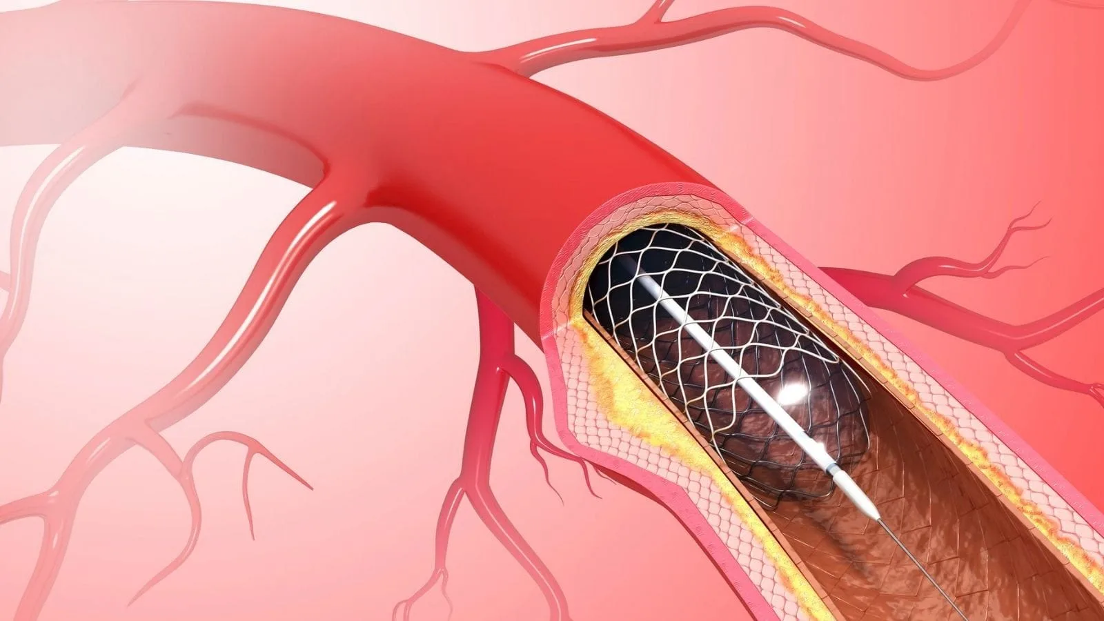
Even when there is no suddenly dislodged clot in the vessel, hard and calcified plaques accumulated in the vessel wall over many years may narrow it so severely that the amount of blood passing through becomes as thin as a thread. In this condition, the tissues in the region continuously suffer from oxygen deprivation, and recurrent minor neurological crises begin. Balloon angioplasty and stenting procedures are performed to repair this narrowed sensitive region from within and widen it permanently.
The first and perhaps most important rule to be considered during the interventional procedure is to prevent small pieces of calcium or fat that may break away from the vessel wall while physically widening the stenosis from traveling to deeper parts of the brain. To ensure this vital safety, a very small protective wire filter that opens like an umbrella is placed behind the narrowing immediately before the procedure. After the area is secured, a very small medical balloon is inflated under high pressure in the narrowed problematic region, stretching the vessel wall outward. After the balloon is deflated, metal mesh stents are permanently left inside the vessel so that it does not collapse inward again due to its own elastic structure and the widened area remains permanently open like a tunnel.
How Are Bulges Around the Medulla Oblongata That May Cause Brain Hemorrhage Closed from Inside the Vessel?
Outward balloon-like enlargements that develop due to thinning and severe weakening of the vessel wall over time are called aneurysms. If these aneurysms in the vessels around the brainstem cannot withstand the pressure and rupture, they cause very severe and sudden bleeding between the membranes of the brain, which carries an extremely high risk to life. With advanced closed interventional methods, these ticking time bombs can be safely occluded and closed directly from inside the vessel without opening the skull in any way.
The main purpose of this closed treatment is to permanently stop high-pressure blood from entering the aneurysm sac and completely eliminate the tension inside the sac, thereby removing the risk of rupture for life. Highly advanced medical technologies designed according to vascular anatomy are used to accomplish this difficult task.
The main treatment materials used include:
- Platinum coils
- Flow-diverting stents
- Liquid embolic agents
- Covered stents
How Are Abnormal Vascular Malformations Around the Medulla Oblongata Treated Without the Need for Open Surgery?
Abnormal vascular malformations that may be present from birth or develop later in life due to structural defects create a high-pressure and extremely dangerous network in the most sensitive areas of the brain. These abnormal formations, in which arteries and veins are directly connected without the balancing capillary vessels between them, carry a very high potential for bleeding. They may also continuously enlarge and exert physical pressure on critical tissues such as the brainstem. Removing these vascular tangles with open surgical methods is an extremely bloody procedure with a high risk of tissue damage.
In closed interventional radiological techniques, the exact center of this highly complex vascular malformation is reached directly using very thin flexible tubes. Once the correct point is reached, specially manufactured liquid adhesive agents that rapidly freeze and harden upon contact with blood are injected into the vessel. This powerful liquid substance spreads throughout and fills all the spaces inside the vascular malformation, freezing within seconds and turning the dangerous area almost into a lifeless stone. Since the blood flow directed into the malformation is instantly and completely stopped, the risk of bleeding is eliminated, and the pressure sensation caused by the swollen vessels on the surrounding tissues also disappears completely over time.
What Kind of Follow-Up Process Awaits Patients After Closed Procedures Performed on the Vessels of the Medulla Oblongata?
The fact that all these closed and minimally damaging interventions do not create major trauma in the tissues makes the recovery process much less painful, more comfortable, and faster than expected for the treated person. However, the care shown by the patient during the return home and subsequent recovery period is just as important to success as the flawless completion of the procedure in the angiography room. After the procedure is completed through the groin or wrist and the instruments are removed, a tight bandage is wrapped around the area where the needle entered, or highly specialized medical vascular closure devices are used to prevent later bleeding or swelling in the artery.
It is extremely important to follow specific rules precisely in order to safely get through the first days after the procedure, during which the body adapts to the new blood flow. Managing the discharge process in accordance with the doctor’s recommendations is the only and most certain way to ensure lifelong success of the delicate stent or platinum procedures applied to the brain vessels.
The steps patients should pay attention to after the procedure include:
- Consuming plenty of fluids
- Regular rest
- Taking medications without interruption
- Regular radiological checkups
- Avoiding strenuous exercise











Vaka Örnekleri