


Cancer is a serious disease that occurs when cells grow uncontrollably in an abnormal way and spread to surrounding tissues. It arises as a result of genetic mutations and, if left untreated, can metastasize and affect vital organs. Early diagnosis increases the success of treatment.
Cancer symptoms usually vary depending on the type of tumor and the organ in which it is located. Common symptoms include unexplained weight loss, persistent fatigue, formation of lumps, bleeding, and prolonged cough. Symptoms should be evaluated without being ignored.
Among the causes of cancer are genetic predisposition, smoking and alcohol use, radiation, certain infections, and environmental toxins. In addition, unhealthy diet, obesity, and physical inactivity are also factors that increase the risk. Prevention is possible by reducing risk factors.
In the diagnostic process, methods such as imaging techniques, laboratory tests, and biopsy are used. After determining the stage and type of cancer, treatment options such as surgery, chemotherapy, radiotherapy, or immunotherapy can be applied. Personalized treatment plans are important.
What Is Cancer and How Does It Develop at the Cellular Level?
To understand cancer, it is necessary to first look at the functioning of a healthy body. The order in our body is maintained by the programmed cell death we call “apoptosis.” In other words, every cell that ages or loses its function quietly withdraws from the stage to make room for a new one. Cancer, biologically, is a disease of resisting this death command. Damage occurring in the genetic material disrupts the cell’s braking mechanism. Like a car with failed brakes speeding downhill, these cells begin to divide continuously.
This process does not occur overnight. It is the result of an accumulation over years. In the first stage, this genetic deterioration that begins in a single cell is also transmitted to the copies of that cell. Over time, this faulty cell population transforms into an organized structure capable of establishing its own nutritional network. Cancer tissue deceives the body into building new vascular pathways to carry oxygen and nutrients to itself. This condition is called “angiogenesis.” Many of the modern interventional treatments we apply are based on cutting off this logistical support of cancer.
What Are the Differences Between a Cancer Cell and a Normal Cell?
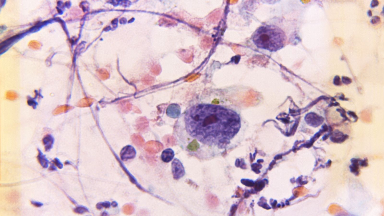
When we examine a cancer cell under a microscope or observe its behavior, we see how much it differs from a normal body cell and how it almost turns into a selfish organism focused on survival. Understanding these differences helps us understand why treatments are very successful in some patients and more challenging in others. Normal cells are social entities; they stop when they touch a neighboring cell and wait their turn. A cancer cell, on the other hand, is antisocial; it listens to no one and grows by stepping over others.
The main features that distinguish cancer cells from healthy cells are:
- Ability to grow uncontrollably
- Immortality
- Producing its own growth signals
- Insensitivity to signals
- Ability to metastasize
- Structural irregularity
- Different energy metabolism
- Ability to form new blood vessels
The item “different energy metabolism” in this list is quite interesting. Cancer cells consume much more sugar (glucose) than normal cells. In fact, they need approximately 100 times more sugar than normal. This greed is the answer to why cancerous tissues shine brightly in imaging methods such as PET-CT; because they immediately absorb the labeled sugar we give them.
What Are the Causes and Risk Factors That Trigger Cancer?
My patients often ask, “Why me?” or “Why did this disease find us?” Unfortunately, the answer to this question is not tied to a single cause. Cancer is an unfortunate intersection of our genetic inheritance with our lifestyle and environmental exposures. A small portion of cases occurs due to defective genes passed down from parents (for example, BRCA mutations). In these individuals, the cell repair mechanism is inherently weak. However, the vast majority of cancers arise from the accumulation of acquired damage over time.
Scientifically proven risk factors that prepare the ground for cancer are:
- Tobacco use
- Alcohol consumption
- Obesity
- Sedentary lifestyle
- Unbalanced diet
- Ultraviolet rays
- Radiation exposure
- Industrial chemicals
- Air pollution
- Chronic infections
- Hormonal imbalances
- Advanced age
- Family history
- Immune system problems
Here, it is especially necessary to emphasize smoking and obesity. Tobacco use disrupts cell structure not only in the lungs but throughout the body, from the bladder to the larynx. Obesity, on the other hand, creates a constant state of inflammation in the body, making it easier for cells to mutate. The increase in cancer risk with age is also related to the gradual fatigue of the body’s repair mechanisms and their tendency to overlook errors.
What Symptoms Does the Body Give as a Warning for Cancer?
Cancer is an insidious disease, but the body often sends us signals that something is wrong. The important thing is not to ignore these signals by saying “it’s just fatigue” or “I must have caught a cold.” Although symptoms vary depending on the location and size of the tumor, some general changes should be considered warning signs. Signals caught at an early stage dramatically increase the success of treatment.
General symptoms to watch out for include:
- Rapid weight loss
- Extreme fatigue
- Night sweats
- Persistent fever
- Unexplained pain
- Loss of appetite
These are the body’s general alarm state. There are also more local symptoms specific to the organ where the cancer is located. For example, a non-healing mouth sore, a mole on the skin changing shape or color, sudden changes in urinary or bowel habits, persistent cough or hoarseness, palpable lumps and swellings are the body’s way of saying “look here.” Especially conditions such as post-menopausal bleeding or seeing blood in the stool are red alarms that require immediate medical attention.
Why Are Interventional Methods and Biopsy Important in the Diagnostic Process?
When you consult a physician with a suspicion of a mass, the process usually begins with imaging methods such as ultrasound, computed tomography (CT), or MRI. These devices provide us with a map of the inside of the body, showing where the mass is and its size. However, there is a very critical fact: imaging methods only tell us that there is a “lesion” there, but they cannot name it. Is what we see an infection, a benign nodule, or an aggressive cancer? The only and definitive way to understand this is to take a sample from that tissue and examine it under a microscope. This procedure is called a “biopsy.”
In the past, patients had to go to the operating room and undergo incisions even for a biopsy. Today, thanks to “Interventional Radiology,” this process has completely changed. We use imaging devices as guidance to reach the suspicious tissue through needle punctures, without making any incision in the body. This method provides great comfort and safety for our patients. Treatment cannot be planned without a diagnosis; because cancer is not a single disease. Even cancers arising in the same organ can have dozens of different subtypes, each with different medications and treatments. Biopsy is the compass that draws this roadmap for us.
What Are the Differences Between Fine Needle and Core Needle Biopsy?
When it comes to biopsy, the most common concern of my patients is “Will it hurt?” or “Will there be a cut?” In biopsies we perform in interventional radiology, no scalpel or surgical knife is used. Procedures are performed under local anesthesia, meaning only that area is numbed, and the patient is awake during the procedure but does not feel pain. We have two main biopsy methods, and which one to use is decided according to the location and type of the mass.
These methods are:
- Fine Needle Aspiration Biopsy
- Tru-Cut (Core Needle) Biopsy
Fine needle biopsy, as the name suggests, is performed with tips even thinner than the needles used for drawing blood. We usually prefer it for thyroid nodules or superficial lymph nodes. We enter the mass with the needle and aspirate the cells by creating suction. It is a very gentle procedure, and the risk of bleeding is almost negligible.
However, in today’s oncology, the real gold standard is the core needle biopsy, which we call “Tru-Cut” or “Core” biopsy. We use this method for masses in organs such as the breast, liver, lung, kidney, and prostate. These needles, which have a special gun mechanism, take a thread-like piece of tissue of millimetric thickness (tissue strip) from inside the tissue. Why do we need this? Because it is not enough to see only the cells; we need to understand the architecture of the tissue, the arrangement of the cells, and the genetic profile of the tumor. Genetic tests that determine whether smart drugs or immunotherapy will work can only be performed on these tissue samples obtained with a core needle. It has an accuracy rate of over 95% and is the most reliable way of diagnosis.
What Does Interventional Radiology Do in Cancer Treatment?
After passing the diagnostic stage, interventional radiology has now become a strong partner to surgery and oncology in the treatment phase. We can define our field as “the technological face of medicine.” The essence of what we do is to reach tumors located in the deepest parts of the body and destroy them under the guidance of imaging devices (ultrasound, angiography, tomography), without making large surgical incisions or putting the patient under deep anesthesia.
The advantages of this “minimally invasive” approach for patients are very clear. First of all, the recovery period is very short. While patients stay in the hospital for days after open surgeries, in our procedures they can usually come in the morning and go home in the evening. The risk of infection is minimal. In addition, cancer patients are often elderly or have comorbidities (heart disease, diabetes, hypertension). For these patients, undergoing general anesthesia can be a significant risk. Since most interventional procedures are performed under local anesthesia and mild sedation (calming), this risk is eliminated. With the philosophy of “no incision, no stitches, no long hospitalization,” we aim to preserve the patient’s quality of life while targeting cancer.
Is It Possible to Destroy a Tumor by Burning or Freezing Without Surgery?
Yes, it is possible, and these methods have become part of standard treatments today. In this group, which we medically call “Percutaneous Ablation,” we destroy the tumor in place using physical methods. We achieve very successful results especially in liver, kidney, lung, and bone tumors in patients who are not suitable for surgery or do not want to undergo surgery.
The main ablation methods applied are:
- Radiofrequency Ablation
- Microwave Ablation
- Cryoablation
- NanoKnife
Radiofrequency (RFA) and Microwave (MWA) methods work on the principle of “destroying by heating.” Under the guidance of tomography or ultrasound, we place a special needle directly into the center of the tumor. The energy sent from the tip of this needle causes molecular vibration in the tumor tissue, generating high heat (between 60-100 degrees). This heat instantly kills tumor cells by cooking them. During the procedure, we make millimetric calculations to avoid damaging surrounding tissues. Microwave ablation provides a faster and stronger effect in larger tumors.
Cryoablation, on the other hand, works with the opposite logic; we destroy the tumor by freezing it. Thanks to gases passing through the needle, the tumor tissue is cooled down to minus 80 degrees. During this process, we observe the formation of an “ice-ball” on the screen that traps the tumor inside. This method provides success equivalent to surgery especially in kidney cancers, and post-procedure pain is much less.
There is also a special technology called “NanoKnife,” which does not use heat. In this method, we apply high-voltage electrical currents to the tumor, creating permanent pores in the cell membranes. The electric current only affects cancer cells; it does not damage critical structures such as vessels, nerves, or bile ducts. Therefore, it can be a life-saving option in pancreatic tumors considered “untouchable” or in liver tumors located in very risky areas.
How Are Intra-Arterial Radiation Therapy and Chemotherapy Applied?
Cancer tissue requires much more nutrients than normal tissue and feeds on them through arteries. By using this weak point, we can strike the tumor “from within.” We enter through the artery in the groin and advance with hair-thin catheters into the liver, directly to the entrance of the vessel feeding the tumor. These procedures are performed in the angiography unit and are called “Transarterial Treatments.”
The types of these methods are:
- Chemoembolization
- Radioembolization
In the chemoembolization (TACE) procedure, we inject a high dose of chemotherapy drug directly into the tumor’s vessel. Along with the drug, we also give micro particles that block the vessel. Thus, the tumor is both subjected to a drug bombardment and deprived of its nutrient supply. Since we deliver the drug only to the tumor instead of the entire body through the bloodstream, side effects such as hair loss or severe nausea are much less common.
Radioembolization (TARE), also known as “radioactive microsphere therapy” among the public, is a much more sophisticated method. Millions of microscopic spheres carrying radioactive material (Yttrium-90) are injected into the tumor vessel again using the angiographic method. These “smart beads” settle inside the tumor and destroy it by emitting radiation from within. While there is a dose limitation in external radiation therapies (radiotherapy) to protect healthy tissue, in this method we can deliver very high and lethal doses directly to the tumor bed. Since the effect area is millimetric, the healthy liver tissue is not damaged. It is a very effective weapon especially in primary liver cancers and metastases that have spread to the liver from other organs.
What Are Supportive Treatments That Increase the Comfort of Cancer Patients?
Interventional radiology not only destroys the tumor but also intervenes to alleviate the difficulties patients face during the treatment process. Cancer treatment is a long-term marathon, and maintaining a high quality of life during this process is very important in terms of morale and physical resilience.
The supportive procedures we frequently perform are:
- Port catheter placement
- Biliary drainage
- Nephrostomy placement
- Ascites fluid drainage
- Pain treatments
Chemotherapy drugs can irritate the veins. Trying to open a vein each time can become a torment for the patient. Therefore, we place a small reservoir called a “Port” under the skin, usually in the chest area. This procedure takes 15-20 minutes. The patient can have all blood tests and chemotherapy administered through this port for months or even years without any pain.
Sometimes tumors can grow and block the bile ducts or urinary channels. In this case, the patient develops jaundice, severe itching, or kidney failure. Without the need for surgery, we enter through the skin with very thin catheters and open these blocked pathways. If necessary, we place permanent metal cages (stents) inside to ensure the normal flow of bile or urine. This is an intervention that saves the patient from intoxication and provides immediate relief. Similarly, draining fluid (ascites) accumulated in the abdomen or performing nerve blocks for cancer-related pain are also practices that make our patients’ lives easier.
Frequently Asked Questions
What is cancer and how does it develop in the body?
Cancer is a disease in which abnormal cells grow uncontrollably and may invade nearby tissues or spread to distant organs. It develops when genetic changes disrupt the normal processes that regulate cell growth and death.
What are the most common early symptoms of cancer that should not be ignored?
Early symptoms vary by cancer type but may include unexplained weight loss, persistent fatigue, unusual lumps, prolonged pain, abnormal bleeding, chronic cough, or changes in bowel, bladder, or skin appearance.
What causes cancer and how do genetic and environmental factors contribute?
Cancer can result from a combination of inherited genetic mutations and environmental exposures. Factors such as tobacco use, radiation, infections, unhealthy lifestyle habits, and certain chemicals may increase risk.
Can cancer develop without causing symptoms in the early stages?
Yes, many cancers may not produce noticeable symptoms initially. This is why screening programs and routine medical evaluations are important, especially for individuals with known risk factors or family history.
Which lifestyle factors are most strongly linked to an increased risk of cancer?
Smoking, excessive alcohol consumption, obesity, poor diet, physical inactivity, excessive sun exposure, and certain occupational exposures are among the most significant lifestyle-related cancer risk factors.
How do doctors determine whether symptoms are caused by cancer?
Diagnosis may involve physical examinations, blood tests, imaging studies such as CT or MRI scans, endoscopic procedures, and biopsies. A tissue biopsy is often required to confirm a cancer diagnosis.
Can cancer spread to other organs and what symptoms might this cause?
Yes, cancer can metastasize to distant organs through the bloodstream or lymphatic system. Symptoms depend on the affected area and may include pain, breathing difficulties, neurological changes, or organ dysfunction.
What are the most effective ways to reduce the risk of developing cancer?
Risk reduction strategies include avoiding tobacco, maintaining a healthy weight, exercising regularly, limiting alcohol intake, protecting skin from excessive sun exposure, and participating in recommended screening programs.
Are all tumors cancerous or can some be noncancerous?
Not all tumors are cancerous. Benign tumors do not invade surrounding tissues or spread to distant organs, while malignant tumors are cancerous and have the potential to grow aggressively and metastasize.
When should persistent symptoms raise concern about the possibility of cancer?
Medical evaluation is recommended when symptoms are unexplained, persistent, worsening, or accompanied by warning signs such as weight loss, abnormal bleeding, a growing lump, or prolonged fatigue.

Prof. Dr. Özgür Kılıçkesmez is one of Türkiye’s leading interventional radiology and neuroradiology specialists, focusing on minimally invasive endovascular treatments. His primary areas of expertise include aneurysm and stroke treatments, embolization procedures, non-surgical treatments of thyroid and parathyroid diseases, interventional oncology, and peripheral vascular interventions. He currently serves as the Head of the Interventional Radiology and Neuroradiology Department at Istanbul Memorial Göztepe Hospital.
Graduating from Istanbul University Cerrahpaşa Faculty of Medicine in 1997, Prof. Dr. Kılıçkesmez received advanced training in interventional radiology and oncology as a Clinical Fellow at London Guy’s & St Thomas’ and King’s College Hospital between 2012 and 2013. He earned the title of Professor in 2020 and, in the same year, became the Founding Interventional Radiologist at SBÜ Çam and Sakura City Hospital. He also holds the EBIR (European Board of Interventional Radiology) certification.
Prof. Dr. Kılıçkesmez has 94 international and 40 national publications and over 2500 citations. He serves as a reviewer for prestigious journals such as the American Journal of Roentgenology (AJR) and Diagnostic & Interventional Radiology. With his clinical and academic expertise, he is a nationally and internationally recognized authority in stroke treatment, brain aneurysm, embolization, tumor ablation, and minimally invasive treatments of thyroid/parathyroid diseases.




Vaka Örnekleri