


Neck pain is a musculoskeletal condition characterized by discomfort, stiffness, or limited mobility in the cervical spine region, often resulting from muscle strain, poor posture, degenerative changes, or underlying medical conditions affecting soft tissues and vertebral structures.
Chronic neck pain symptoms and causes include prolonged sedentary behavior, improper ergonomic positioning, cervical disc degeneration, and stress-related muscle tension, all of which contribute to persistent discomfort, reduced range of motion, and potential nerve compression in advanced cases.
Acute neck pain treatment methods focus on pain management through physical therapy, anti-inflammatory medications, posture correction, and controlled exercises designed to restore cervical alignment and improve muscle strength while preventing further strain or injury.
Neck pain prevention and risk factors involve maintaining proper posture, ergonomic workspace adjustments, regular stretching routines, and early intervention strategies to minimize the progression of cervical spine disorders and reduce the likelihood of chronic pain development.
What Causes Neck Pain and What Are Its Anatomical Foundations?
To understand the neck region, one must first imagine the load it carries. We’re talking about a structure that holds our head—approximately 4–5 kilograms—upright throughout the day, enables us to turn it in all directions, and protects vital nerve highways running from the brain to the body. The uppermost seven vertebrae of the 33 in the spinal system form the cervical spine or neck region. Between these bones are discs that facilitate movement and function like shock absorbers.
When we trace the origin of neck pain, we typically encounter mechanical strain or inflammation developed within the body. In today’s world, the biggest enemies are immobility and poor posture habits. When we tilt our heads forward to look at a phone screen for hours, the load on the cervical vertebrae multiplies. This causes excessive tension in the neck muscles, ligaments, and fascia.
However, muscle fatigue isn’t the only culprit. When structural deterioration comes into play, things get more complex. Especially with aging, a natural wear-and-tear process begins in the spine. Over time, discs lose water, become less elastic, and reduce in height. When joint degeneration is added to this, pain becomes inevitable.
The most common anatomical causes of neck pain include:
- Poor posture
- Overuse of neck muscles
- Injuries due to sudden movements
- Cervical disc herniation
- Cervical spondylosis
- Facet joint degeneration
- Spinal canal stenosis
- Traumatic injuries
In addition to these, more rarely, persistent and stubborn pain may be due to tumors, vascular issues, or rheumatic diseases. Therefore, for pain that doesn’t subside with rest, wakes you up at night, or progressively worsens, a detailed examination is essential.
What Is the Difference Between Axial and Radicular Neck Pain?
As physicians, the nature of the pain tells us a lot. Whether the pain remains localized in the neck or radiates to other areas is the most fundamental distinction guiding our treatment path. In medical literature, we generally classify these pains into three main groups: axial, radicular, and myelopathic. This distinction determines the type of intervention to be made.
Axial pain is typically described by patients as “a heaviness at the base of my neck” or “a dull ache spreading from my neck to my shoulders.” This type of pain doesn’t radiate to the arm or hand. The usual cause lies in joint degeneration between the cervical vertebrae, disc wear, or muscle tension. There is no direct pressure on the nerve root—this is a problem primarily with the spine’s mechanical components.
Radicular pain, however, presents a completely different picture. Patients often describe it as “like an electric shock going down my arm” or “it feels like a vein is being pulled.” Here, the issue is compression of a nerve root exiting the spinal cord en route to the arm. This compression is usually due to a herniated disc or narrowed bony canal. The pinched nerve swells, causing severe, burning pain.
Myelopathic pain, on the other hand, is a more serious condition involving direct compression of the spinal cord. It’s not just pain—it also presents with neurological deficits like clumsiness in the hands, inability to button shirts, or leg weakness.
The most prominent features of axial pain include:
- Localized pain at the back of the neck
- Pain radiating to the shoulder girdle
- Morning stiffness
- Restricted neck movement
- Headaches
The most prominent features of radicular pain include:
- Sharp pain radiating to the arm
- Numbness in the fingers
- Tingling sensation
- Weakness in arm muscles
- Loss of reflexes
What Imaging Techniques Are Used to Diagnose Disc Herniation and Osteoarthritis?
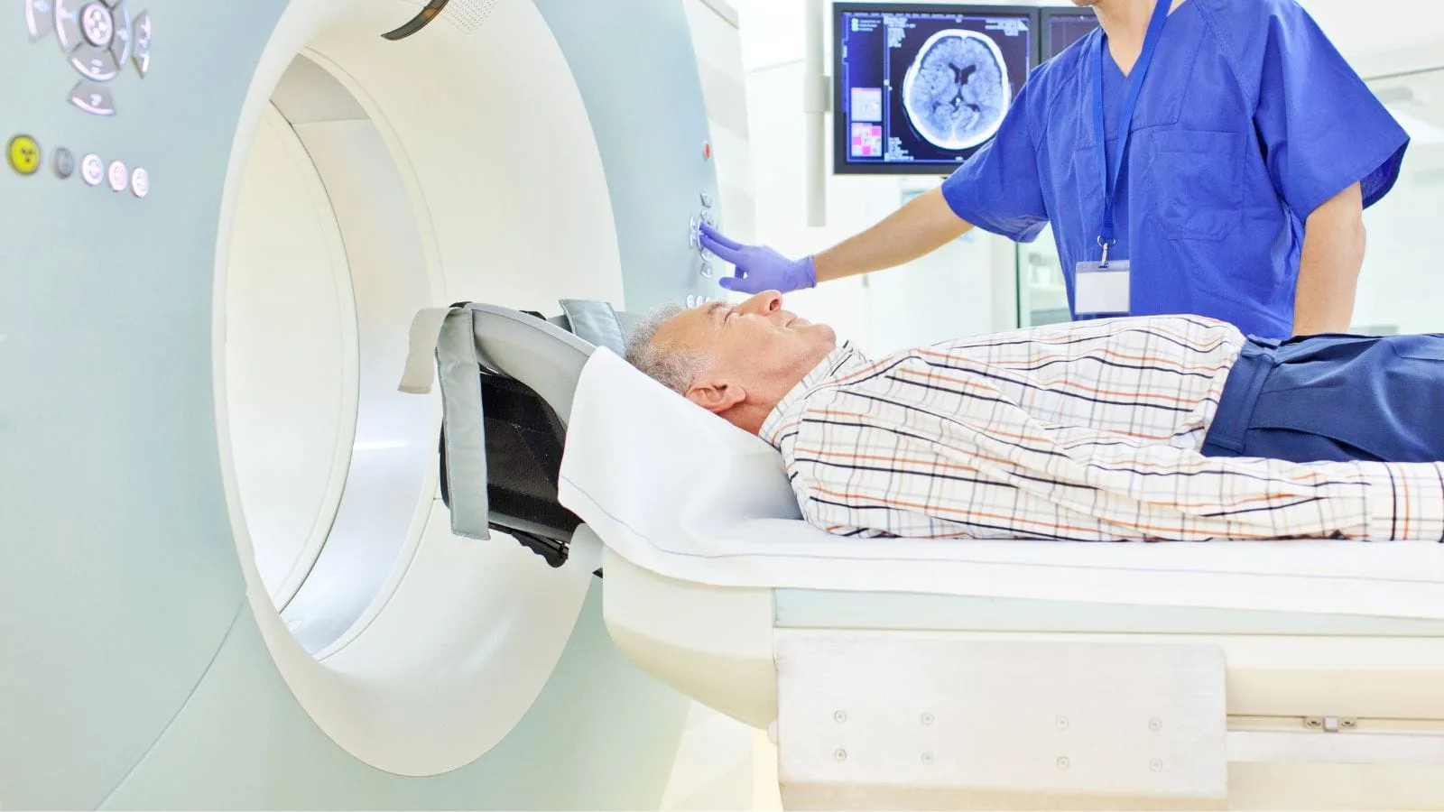
The greatest strength of modern medicine—and especially interventional radiology—is the principle of “do not treat what you cannot see.” After listening to the patient’s complaints, we use advanced imaging techniques to identify the pain source. At this point, Magnetic Resonance Imaging (MRI) becomes our indispensable guide.
Unlike X-rays, MRI reveals not only bones but also discs, nerves, the spinal cord, and soft tissues in great detail. With MRI, we can clearly see the level of a cervical disc herniation, how much pressure it puts on the nerve, the degree of facet joint degeneration, or any spinal canal narrowing. It also helps us distinguish pain-causing conditions like tumors, infections, or rheumatic processes.
For interventional radiologists, MRI is not just a diagnostic tool—it’s a map. We plan the location of needle-based treatments and identify the target nerve root using these images. However, a crucial detail is this: an MRI image isn’t everything. Sometimes a herniated disc may appear severe on MRI yet cause no pain, while a small issue may cause intense pain. Therefore, imaging must always be correlated with the patient’s symptoms.
How Is the Pain Source Definitively Identified with Diagnostic Blocks?
Imaging methods show us anatomical abnormalities, but they do not 100% confirm that the pain originates from those abnormalities. Especially in complex cases where the patient has both a herniated disc and osteoarthritis, identifying the actual pain source is critical to treatment success. This is where the method known as “diagnostic blocks” comes into play.
This procedure is essentially a temporary test. We inject a very low dose of local anesthetic into the suspected pain source (e.g., a specific nerve root or joint). If the patient experiences a more than 50% reduction or complete disappearance of pain immediately after the procedure, we are confident: this is the source of the pain. This method eliminates trial-and-error and ensures that the permanent treatment targets the correct area.
Advantages of diagnostic blocks include:
- Precise identification of the pain source
- Prediction of treatment success
- Prevention of unnecessary surgeries
- Elimination of the risk of treating the wrong area
- Providing prognostic information to the patient
How Is the Targeted “Pinpoint” Treatment Applied in Cervical Disc Herniation?
For neck and arm pain that persists despite medication, rest, and physical therapy, one of our most effective non-surgical tools is the Transforaminal Epidural Injection, commonly referred to as “Pinpoint” therapy. This method is particularly effective in nerve compression (radiculopathy) due to cervical disc herniation.
The logic is this: medications taken orally or via injection into the hip circulate throughout the entire body and reach the problematic area in minimal concentrations. But with pinpoint therapy, we deliver the medication directly to the problem’s core. Using a fine needle, we enter the narrow channels of the spine called foramina, where nerves exit, and reach the compressed, swollen nerve root.
The injected mixture typically includes a long-acting corticosteroid and a local anesthetic. The purpose of the steroid is not bodybuilding—it powerfully reduces the inflammation in that area and resolves the nerve edema. When the swelling subsides, the nerve relaxes, and the pain cycle is broken. This treatment does not mechanically eliminate the herniation but removes the chemical and physical irritation it causes, promoting healing.
What Is Radiofrequency Ablation Therapy for Cervical Osteoarthritis?
If the patient’s pain isn’t from a herniated disc but from degeneration of the facet joints located at the back of the cervical vertebrae, our treatment approach changes. These patients usually struggle to turn their necks and have predominant neck pain. In such cases—called facet syndrome—we use Radiofrequency Ablation (RFA), also known as “Nerve Burning.”
While the name may sound intimidating, the nerve is not charred like coal. In this procedure, we target the small nerve endings that carry pain signals from the joint to the brain. With a special needle, we approach these nerves and deliver controlled thermal energy via radiofrequency waves. Typically, a temperature of around 80°C is applied for 90 seconds, which disables the tiny nerve fibers’ ability to transmit pain.
Thanks to this procedure, even though the joint issue remains, the brain no longer receives the “pain” signal, so the patient does not feel pain. It’s similar to numbing a tooth before a filling, but the effect lasts for months or even years, not hours. Nerves may regenerate over time, so the pain might return years later—but the procedure can be safely repeated.
Conditions treated with radiofrequency ablation include:
- Chronic neck pain
- Facet joint osteoarthritis
- Pain following whiplash injuries
- Cervicogenic headaches
- Shoulder girdle pain
How Is Safety Ensured in Interventional Procedures and Why Is Fluoroscopy Used?
The neck is one of the body’s most complex and risky junctions. Carotid arteries, the spinal cord, the trachea, and vital nerves are all packed into a very tight space. Therefore, injections into the neck should never be done blindly or based solely on palpation. The most important rule in interventional radiology is: never inject medication where you cannot see the needle tip.
To ensure safety, we perform all procedures under fluoroscopy, a type of real-time X-ray. This device shows us the bones and needle progression on the screen. But seeing bones alone is not enough. Is the needle in the right place? Is it inside a blood vessel? Is the medication spreading around the nerve or going elsewhere?
To answer these questions, we use contrast agents (radio-opaque dye). When we inject this dye, we see a black spread on the screen and confirm whether the drug is correctly enveloping the target nerve root. This step is vital for the accuracy and safety of the procedure.
Safety steps during procedures include:
- Continuous fluoroscopic imaging
- Verification with contrast dye
- Neurophysiological stimulation
- Monitoring of vital signs
- Sterile operating room conditions
Why Is Control with Nerve Stimulation Performed?
Especially in radiofrequency (burning) procedures, we use technology to further enhance safety. After placing the needle—and before delivering medication or performing ablation—we send low-voltage electrical currents from the needle tip. This is called neurophysiological stimulation.
It serves two purposes. First, are we next to the correct nerve? When we apply low current, we want the patient to feel tingling or a tapping sensation where they normally feel pain. This confirms, “yes, we’re on target.” Second, and more importantly, are we in a safe location? When we use a different frequency, we want no movement in the arm or hand. If the arm moves, the needle is too close to a motor nerve, and treating there is dangerous. We slightly reposition the needle and retest. This dual control mechanism is essential to prevent unwanted nerve injury.
Is General Anesthesia Used During These Procedures?
One of the most frequently asked questions is whether the patient will be put to sleep. One of the greatest advantages of these minimally invasive procedures is that they do not require general anesthesia. The patient’s awareness and ability to communicate with us during the procedure are part of the safety protocol. For example, we might ask, “Where is your pain now?” or “Do you feel an electric shock in your arm?” and adjust accordingly.
That said, patient comfort is very important. To prevent pain or anxiety, we administer sedation through an IV. Additionally, we use local anesthesia (like dentists do) to numb the needle entry site. As a result, the patient doesn’t recall the procedure or only remembers slight pressure—but they do not fall into a deep sleep or require a breathing machine.
What Is the Recovery Process After Treatment and What Should Be Watched For?
The main difference between interventional radiology and surgery is the short recovery time. Since no scalpel is used, muscles are not cut, and no stitches are required, the body recovers very quickly. The procedure usually takes 15 to 45 minutes.
After the procedure, the patient is transferred to the recovery room on a stretcher. A small dressing is applied to the entry site, and a sandbag is placed on top to prevent bleeding. After about four hours of observation, if the patient feels well, walks normally, and has stable vital signs, they are discharged the same day.
Post-procedure at home is generally very comfortable. Rest is advised on the first day, but patients usually return to simple daily activities by the next day. There may be mild soreness at the needle entry site, which is managed with ice packs and simple pain relievers.
With epidural steroid injections, the effect of the corticosteroid may not start immediately. A feeling of fullness or a temporary increase in pain may occur due to the medication’s volume. The true healing effect usually begins on day 3 and reaches its peak in the following weeks.
With radiofrequency procedures, there may be a temporary burning sensation or slight increase in pain for 1–2 weeks due to nerve heating. This is expected and part of the temporary phase before permanent relief sets in.
Key post-procedure precautions include:
- 24 hours of bed rest
- Plenty of fluid intake
- Avoid heavy lifting
- Refrain from sudden neck movements
- Watch for signs of infection
- Follow the prescribed timing for starting exercises
Frequently Asked Questions
What is neck pain and how can it affect daily activities?
What are the most common causes of neck pain in adults?
Can poor posture and screen use contribute to chronic neck pain?
What symptoms accompanying neck pain may indicate a more serious condition?
How can cervical disc herniation cause neck pain and related symptoms?
How is the underlying cause of neck pain diagnosed?
What helps relieve neck pain without surgery?
When is surgery considered for patients with neck pain?
Can exercise and physical therapy help prevent recurring neck pain?
What experiences do patients commonly report after successful neck pain treatment?

Prof. Dr. Özgür Kılıçkesmez is one of Türkiye’s leading interventional radiology and neuroradiology specialists, focusing on minimally invasive endovascular treatments. His primary areas of expertise include aneurysm and stroke treatments, embolization procedures, non-surgical treatments of thyroid and parathyroid diseases, interventional oncology, and peripheral vascular interventions. He currently serves as the Head of the Interventional Radiology and Neuroradiology Department at Istanbul Memorial Göztepe Hospital.
Graduating from Istanbul University Cerrahpaşa Faculty of Medicine in 1997, Prof. Dr. Kılıçkesmez received advanced training in interventional radiology and oncology as a Clinical Fellow at London Guy’s & St Thomas’ and King’s College Hospital between 2012 and 2013. He earned the title of Professor in 2020 and, in the same year, became the Founding Interventional Radiologist at SBÜ Çam and Sakura City Hospital. He also holds the EBIR (European Board of Interventional Radiology) certification.
Prof. Dr. Kılıçkesmez has 94 international and 40 national publications and over 2500 citations. He serves as a reviewer for prestigious journals such as the American Journal of Roentgenology (AJR) and Diagnostic & Interventional Radiology. With his clinical and academic expertise, he is a nationally and internationally recognized authority in stroke treatment, brain aneurysm, embolization, tumor ablation, and minimally invasive treatments of thyroid/parathyroid diseases.





Vaka Örnekleri