


Heparin is a powerful anticoagulant drug used to suppress the blood clotting process and prevent thrombus formation. It helps maintain blood flow by preventing clot development within vessels and is administered under medical supervision to provide rapid-acting anticoagulation in acute or high-risk situations.
Clinical conditions in which heparin therapy is used include deep vein thrombosis, pulmonary embolism, acute coronary syndrome, and reducing clot risk after certain surgical procedures. It is also preferred as a preventive treatment for clot formation in dialysis applications and to maintain the patency of intravascular catheters.
Heparin administration methods and dosing vary depending on the patient’s clinical condition and treatment purpose. The drug is usually administered as an intravenous infusion or subcutaneous injection. The dose is adjusted based on the patient’s weight, coagulation tests, and comorbidities, and laboratory values are closely monitored throughout treatment.
Monitoring and safety are of great importance in heparin use because the risk of bleeding may increase during treatment. Coagulation tests such as activated partial thromboplastin time are regularly checked. In addition, clinical evaluation is performed for early detection of rare but serious side effects such as heparin-induced thrombocytopenia.
| Things You Need to Know | Information |
| What Is Heparin | Heparin is a powerful anticoagulant (blood thinner) that prevents clot formation and inhibits blood clotting within vessels. It is a directly acting anticoagulant and is generally administered intravenously or subcutaneously. |
| Mechanism of Action | It activates Antithrombin III and inhibits clotting factors, especially thrombin and factor Xa. Thus, it prevents new clot formation and stops the growth of existing clots. |
| Indications for Use | Treatment and prophylaxis of thromboembolic risk conditions such as deep vein thrombosis (DVT), pulmonary embolism, myocardial infarction, and atrial fibrillation. It is also used to prevent clot formation in interventional procedures such as pre/postoperative settings or dialysis. |
| Method of Administration | Administered as subcutaneous injection or intravenous infusion. In intravenous applications, continuous dosing can be delivered via an infusion pump. |
| Dosage and Monitoring | Treatment doses are determined according to the patient’s weight, condition, and coagulation test results. During heparin therapy, blood clotting time is regularly monitored using the aPTT (activated Partial Thromboplastin Time) test. |
| Main Side Effects | Bleeding (the most serious risk), thrombocytopenia (Heparin-Induced Thrombocytopenia – HIT), osteoporosis (with long-term use), bruising or pain at the injection site. |
| Contraindications | It should not be used in patients with active bleeding, severe thrombocytopenia, heparin allergy, individuals who have undergone certain brain surgeries, and those with uncontrolled hypertension. |
| Use in Pregnancy | Since heparin does not cross the placenta, especially its low molecular weight form (LMWH), it is one of the preferred anticoagulants during pregnancy. |
| Types of Heparin | – Standard (unfractionated) heparin: Requires more frequent monitoring and dose adjustment.- Low molecular weight heparin (LMWH): (e.g., enoxaparin) Provides a more stable effect and requires less monitoring. |
| Antidote in Emergency | Protamine sulfate is used as an antidote in the treatment of excessive bleeding caused by heparin. |
What Is Heparin and How Does It Work in Our Body?
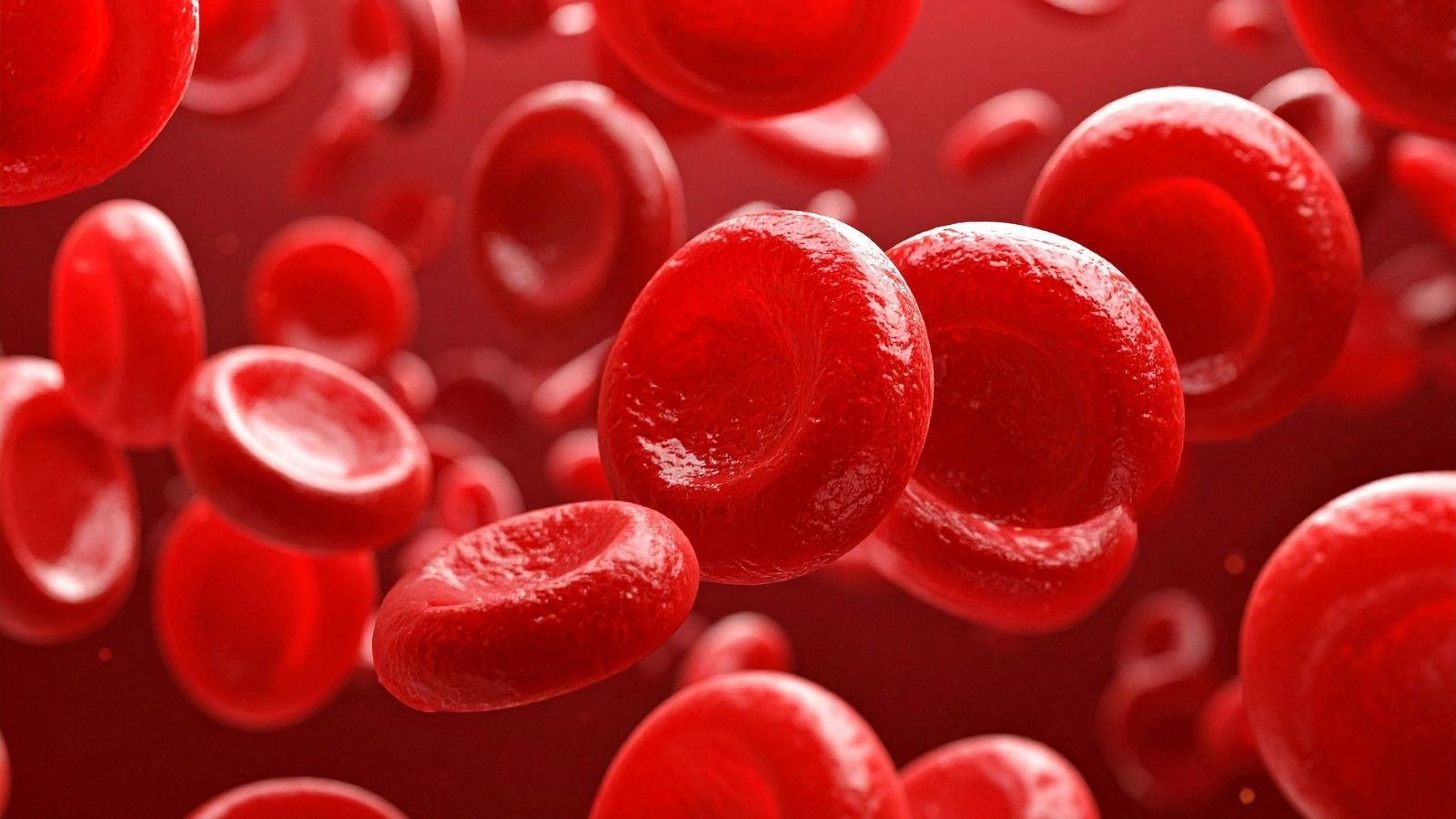
Blood clotting is actually one of the most magnificent and vital defense mechanisms our body possesses. When a small cut occurs on your finger, small blood components called “platelets,” which patrol inside blood vessels, immediately rush to that area. These platelets stick to each other and form a physical plug. Then, various proteins circulating in the blood create a network that strengthens this plug and stops the bleeding. In a healthy person, this system should only activate when there is bleeding. However, in some diseases or during medical interventions within the vessel, this system may mistakenly or excessively activate inside the vessel, leading to dangerous blockages that completely stop blood flow.
This is where heparin comes into play as a very powerful regulator. Chemically, it is a highly complex and long-chain molecule. In fact, this substance is naturally produced by certain cells in human and animal bodies and is one of the biological components with the highest negative electrical charge known. When administered externally as a drug in medical treatments, the mechanism of heparin is not to directly thin the blood or dissolve clots. Instead, it works by modifying the function of a natural protein in our body called “Antithrombin,” which is responsible for slowing down clotting.
When the heparin molecule enters the bloodstream, it binds tightly to this natural inhibitory protein. This binding changes the protein’s three-dimensional structure, increasing its ability to capture and neutralize clotting enzymes by a thousandfold. In other words, heparin acts as a catalyst that accelerates the body’s own safety system to an extraordinary speed. Its ability to act within seconds after intravenous administration makes it an indispensable tool in heart attacks, pulmonary embolisms, and high-risk vascular surgeries requiring emergency intervention.
Why Is Heparin So Important in Interventional Radiology Procedures?
Interventional radiology involves modern medical treatments performed by entering the vessels through a small needle puncture instead of making large surgical incisions, guided by imaging devices. During these procedures, access is gained through the patient’s groin, wrist, or neck vessels, navigating through meters-long vascular networks to reach the targeted problematic area. This area may be a blocked leg vein, a brain aneurysm at risk of bleeding, or an abnormal vascular network feeding a tumor.
During these delicate procedures, the medical equipment advanced within the vascular tunnels, although high-tech, are completely foreign substances to the body. The human immune and clotting system perceives a plastic or metal object moving within the vessel as a major threat or a massive wound. If the clotting system is not temporarily suppressed, blood cells begin to adhere to these devices within seconds.
Clots forming on these devices not only block the materials used but can also detach due to blood flow and travel to the fine vessels of vital organs such as the lungs, heart, or brain, causing sudden death or permanent paralysis. Therefore, maintaining blood fluidity during the procedure is essential not only for successful treatment but also for the patient’s survival. Heparin forms an invisible and slippery molecular shield around these medical devices, preventing blood cells from adhering to their surfaces.
The main medical materials used in interventional procedures that create clotting risk include:
- Catheters
- Guidewires
- Metal stents
- Balloons
- Protective sheaths
What Are the Types of Heparin Used in Medical Treatments?
In modern medical practice, two main types of heparin are generally encountered. Although their mechanisms of action in the body are fundamentally similar, there are major differences between them in terms of administration methods, duration of effect, and monitoring requirements. The choice of type in treatment planning depends entirely on the patient’s current condition, whether hospitalization is required, and the nature of the medical procedure to be performed.
The first type is “Standard Heparin,” or medically known as unfractionated heparin. Its molecular structure remains intact, long, and unfragmented in laboratory conditions. Due to this structure, it provides the best results when administered intravenously. Its greatest advantage is that it shows maximum effect within seconds upon entering the bloodstream and is eliminated from the body very quickly. Additionally, in case of a bleeding complication, its effect can be immediately and completely neutralized with a specific antidote. This rapid on-off feature makes it a perfect option for operating rooms and interventional radiology units where millimetric precision is required.
The second type is known as “Low Molecular Weight Heparin.” This drug is obtained by breaking standard heparin into smaller, uniform, and shorter fragments using special enzymes or chemical processes in laboratory conditions. This process completely changes the molecule’s character. Low molecular weight heparins are administered through small subcutaneous injections. Their absorption is slower, allowing their effect to remain stable for much longer periods. They have a predictable effect profile that does not require continuous blood test-based dose adjustments. Their greatest advantage is that patients can easily administer them at home, either by themselves or with the help of a relative, without requiring hospitalization.
Situations in which low molecular weight heparins that can be used at home are preferred include:
- Pregnancy period
- Prolonged bed rest
- After orthopedic surgery
- During cancer treatment
- After long intercontinental flights
What Are the Steps of Heparin Administration in Intravascular Operations?
When an intravascular treatment is planned, heparin administration is never random; it is based on very strict mathematical rules and a careful protocol. As soon as the patient’s vessel is accessed and the main sheath through which all equipment will pass is placed, the body’s clotting alarm is triggered. At this exact moment, the first high dose of heparin, precisely calculated in milligrams or units according to the patient’s weight, is injected directly into the vein. This is called the “initial bolus dose.”
This initial dose rapidly and strongly suppresses the clotting system. However, since the standard form of heparin is cleared from the body quite quickly, its protective effect in the blood gradually decreases as the procedure duration increases. Opening a complex leg vein occlusion or treating a challenging brain aneurysm may take hours. In such long procedures, the blood level is usually remeasured every hour, and smaller maintenance doses are administered to prevent weakening of the protective shield.
In some cases, the problem is already a massive blood clot. For example, in deep vein thrombosis, where the main leg veins are completely filled with clots, special catheters are placed into the clot and clot-dissolving drugs are slowly infused. This process may take more than twenty-four hours. During this long period, heparin is administered continuously into the patient’s vein as a drip infusion using an electronic pump to prevent the clot from spreading or new clots from forming.
Why Does Heparin Require Extra Sensitivity in Interventions on Brain Vessels?
Among all vascular networks in the body, the most delicate and high-risk area for intervention is undoubtedly the brain vessels. In these procedures, known as neuroendovascular interventions, very fine vascular tunnels extending into the depths of the brain within the skull are accessed. Brain tissue is the most sensitive tissue in the human body to oxygen deprivation and circulation disorders. Even a few minutes of interrupted blood flow can cause irreversible death of millions of brain cells.
A millimetric clot formed during a procedure in the leg or arm vessels may often be tolerated without causing major problems or may only result in mild pain in that region. However, even a tiny clot in brain vessels, barely visible to the eye, can lead directly to a stroke, meaning paralysis, with catastrophic consequences. This fact necessitates a much more aggressive anticoagulation strategy in brain vessel procedures.
However, there is a very delicate balance to be maintained. Brain vessels are structurally much thinner and more fragile than other vessels in the body. Excessive thinning of the blood to prevent clotting may lead to uncontrollable, fatal brain hemorrhage if one of these delicate vessels ruptures. Therefore, during the procedure, the interventional team must walk a very fine line between the risk of stroke and the risk of brain hemorrhage. This perfect balance is achieved through precisely calculated and continuously monitored heparin doses.
The main life-threatening conditions involving brain vessels include:
- Acute stroke
- Brain hemorrhage
- Aneurysms
- Arteriovenous malformations
- Carotid artery stenosis
How Is the Heparin Level in the Patient’s Blood Measured During Surgery?
When a blood-thinning drug is administered, it is vital to know how much effect the drug has on the individual. The human body is not a machine; the same drug dose can cause completely different levels of anticoagulation in two different patients. Even in the same patient, the response to heparin may vary between procedures performed in the morning and in the evening. Therefore, in high-risk intravascular operations, decisions are not made blindly or based on estimation; real-time and continuous measurements are performed.
The most commonly used method for this critical measurement in interventional radiology or angiography units is a special test called Activated Clotting Time. The greatest advantage of this test is that it provides results within seconds directly in the procedure room without needing to send the blood sample to the hospital’s main laboratory. During the procedure, only a few drops of blood are taken from the patient’s catheter, placed into a special tube, and inserted into a small electronic device next to the operating table.
The device initiates the clotting process using a substance added to the blood and measures the time it takes for the blood to clot completely in seconds. In a normal person who has not taken any medication, the clotting time is around one hundred seconds. During procedures such as stenting or ballooning, the target is to increase this time to between two hundred and three hundred seconds with the help of heparin, approximately two and a half times the normal value. The specialist performing the procedure makes precise decisions based on the seconds displayed on the device screen, such as “The protective shield has weakened, let’s add more heparin” or “The clotting level is currently ideal and safe, we can continue the operation.”
How Is the Heparin Lock Method Applied in Long-Term Catheter and Port Maintenance?
One of the main roles of interventional radiologists is not only to open blocked vessels but also to create safe vascular access for patients who will undergo long-term treatments. This includes thick dialysis catheters for patients requiring continuous dialysis due to kidney failure or special port systems placed under the skin for cancer patients who need chemotherapy for months. These devices are usually made of plastic or silicone derivatives and can remain in the patient’s body for months or even years.
The biggest enemy of these devices is the clotting of blood that accumulates inside them during inactive periods when they are not in use, leading to complete blockage of the tube system. Plastic surfaces are always stimulants for clot formation. To prevent these blockages, there is a highly practical and effective method used worldwide as a standard. This method is called a “heparin lock” in medical terminology.
After a chemotherapy session or dialysis procedure is completed, the internal volume of the thin tube portion remaining inside the vessel is calculated with millimetric precision. This internal volume is usually only a few milliliters. This small space is completely filled with a saline solution containing high-concentration heparin, and the device is sealed. This solution does not mix with the patient’s bloodstream; it remains fixed on the inner walls of the tube, creating a mechanical barrier. Weeks later, when the patient returns for another treatment session, the nurse withdraws this heparinized fluid with a syringe before administering new medication and discards it. Thus, the inside of the tube remains completely open, clean, and free of clots before each use.
Problems that may occur in ports that are not regularly maintained include:
- Complete blockage
- Infection
- Treatment delay
- Intravascular inflammation
- Need for complete removal of the device
What Are the Possible Risks and Side Effects of Heparin Therapy?
Like any medical agent that strongly interferes with one of the body’s fundamental defense mechanisms, heparin has risks that must be managed with extreme care and respect. As a natural consequence of artificially stopping blood clotting, the most common and expected side effect of treatment is bleeding. Depending on the drug dose and the patient’s sensitivity, even a small injury anywhere in the body may bleed more and for a longer time than normal. Leakage at needle entry sites or bruising due to large subcutaneous bleeding may be observed. If the patient has a pre-existing sensitive condition such as a stomach ulcer, the risk of serious internal bleeding in these areas must always be considered.
In addition, there is a much rarer but extremely dangerous paradoxical side effect. This condition is known in medical literature as Heparin-Induced Thrombocytopenia. In some patients, the immune system perceives heparin as a dangerous virus or bacteria entering the body and begins to produce special antibodies against it.
These antibodies attack the points where heparin binds to platelets. As a result of this attack, platelets break down and their numbers rapidly decrease; however, more alarmingly, so many clotting substances are released during this breakdown that massive and uncontrolled clots begin to form throughout the body despite the patient receiving strong anticoagulant therapy. This condition usually appears between one week and ten days after starting treatment. It may manifest with sudden bruising in the legs, unexplained shortness of breath, or severe chest pain. If such a condition is suspected, doctors immediately stop heparin and switch to alternative drugs with completely different chemical structures.
Undesirable conditions that may develop due to heparin use include:
- Gum bleeding
- Subcutaneous bruising
- Nosebleeds
- Blood in urine
- Sudden drop in platelet count
How Is the Effect of Heparin Reversed in an Emergency Bleeding Situation?
During interventional radiology procedures or the follow-up period afterward, things may not always go as planned. Emergency situations may occur, such as accidental tearing of a very thin and delicate vessel during the procedure, sudden spikes in blood pressure causing bleeding in a previously unnoticed area, or uncontrollable bleeding from the surgical site. In such moments, one of the most valuable biological properties of heparin comes into play: its ability to be completely and rapidly neutralized.
If it is necessary to urgently restore the patient’s clotting system to its previous strong state, a special antidote called “Protamine Sulfate” is used. Protamine is actually a basic protein derived from the natural protein structures of salmon fish and modified in the laboratory for medical use. Its mechanism of action is based on a very simple chemical principle: the attraction of opposite charges.
As mentioned earlier, heparin molecules carry a very high negative electrical charge. Protamine sulfate, on the other hand, has a strong positive electrical charge. When this antidote is administered into the patient’s vein, it rapidly binds to the negatively charged heparin molecules in the circulation like powerful magnets. As a result of this binding, the two molecules stick together and form a stable salt-like structure, losing their effect and entering the process of elimination from the body. This process is so rapid and effective that the clotting mechanism, which had been suppressed, can return to its normal function within minutes in a bleeding patient. However, the administration of this antidote must also be done slowly, carefully, and with controlled infusion; otherwise, it may cause other serious problems such as sudden drops in blood pressure and heart rhythm disturbances.
How Should Heparin Be Used During Pregnancy and With Other Medications?
The safe management of heparin therapy depends not only on administering the correct dose but also on considering the patient’s overall health status and all other factors in daily life. Especially in elderly individuals and those with chronic diseases, the use of multiple medications simultaneously is very common. If the patient is already taking strong painkillers for rheumatic conditions or other blood-thinning tablets that prevent platelets from sticking together due to heart diseases, the bleeding effect of heparin in the body increases synergistically. If such drug interactions are not controlled, life-threatening internal bleeding may occur in organs such as the stomach or intestines.
On the other hand, one of the most specific and sensitive patient groups in which heparin is used is expectant mothers. Pregnancy is a period when the blood in a woman’s body naturally becomes more prone to clotting in preparation for childbirth. This significantly increases the risk of clot formation, especially in the leg veins. However, administering medication to a pregnant woman is always challenging because most chemicals cross the placental barrier and enter the baby’s circulation, potentially harming its development.
At this point, heparin offers a unique advantage due to its large molecular structure. Heparin and low molecular weight heparin molecules are physically so large that it is biologically impossible for them to pass through the microscopic pores of the placenta and reach the baby. This feature makes them the safest and gold-standard option in the treatment and prevention of clotting disorders during pregnancy. They do not harm the baby’s development in any way. Similarly, due to their large size, they cannot pass into breast milk, allowing them to be used safely during the breastfeeding period without concern.
Things to be cautious about when using heparin together due to increased bleeding risk include:
- Aspirin
- Rheumatism medications
- Other blood thinners
- High-dose corticosteroids
- Platelet-inhibiting agents
Frequently Asked Questions
What Is Heparin and how does it prevent blood clot formation?
Heparin is an anticoagulant medication that helps prevent the formation and growth of blood clots. It works by enhancing the activity of natural proteins that regulate the blood-clotting process and maintain healthy circulation.
What Is Heparin and in which medical situations is it commonly used?
Heparin is frequently used to prevent or treat deep vein thrombosis, pulmonary embolism, heart-related clotting conditions, and clot formation during surgeries, dialysis, or certain interventional procedures.
What Is Heparin and can it be used during hospitalization?
Heparin is commonly administered to hospitalized patients who are at increased risk of developing blood clots due to surgery, prolonged immobility, serious illness, or specific cardiovascular conditions.
How Is Heparin Applied in emergency medical situations?
In emergency settings, heparin may be given intravenously to provide rapid anticoagulation. This approach is often used when immediate clot prevention or treatment is required under close medical supervision.
How Is Heparin Applied for long-term clot prevention?
Depending on the condition being treated, heparin may be administered through subcutaneous injections. Treatment duration varies according to the patient’s diagnosis, risk factors, and overall treatment plan.
What side effects can occur during Heparin treatment?
The most common side effect of heparin is bleeding. Patients may also experience bruising at injection sites, allergic reactions, or changes in platelet counts that require monitoring by healthcare professionals.
Who may require special monitoring while receiving Heparin?
Individuals with kidney disease, bleeding disorders, recent surgery, advanced age, or a history of clotting complications may require closer monitoring to ensure safe and effective anticoagulant therapy.
Can Heparin be used during pregnancy when anticoagulation is needed?
Heparin is often considered when anticoagulation is necessary during pregnancy because it does not significantly cross the placenta. Treatment decisions are individualized based on maternal and fetal health considerations.
How do physicians monitor the effectiveness of Heparin therapy?
Doctors may use blood tests such as activated partial thromboplastin time (aPTT) or anti-factor Xa assays to evaluate the medication’s effectiveness and adjust dosing when necessary.
When should medical attention be sought during Heparin treatment?
Patients should seek prompt medical evaluation if they experience unusual bleeding, severe bruising, blood in the urine or stool, significant swelling, or other symptoms that may indicate treatment-related complications.

Prof. Dr. Özgür Kılıçkesmez is one of Türkiye’s leading interventional radiology and neuroradiology specialists, focusing on minimally invasive endovascular treatments. His primary areas of expertise include aneurysm and stroke treatments, embolization procedures, non-surgical treatments of thyroid and parathyroid diseases, interventional oncology, and peripheral vascular interventions. He currently serves as the Head of the Interventional Radiology and Neuroradiology Department at Istanbul Memorial Göztepe Hospital.
Graduating from Istanbul University Cerrahpaşa Faculty of Medicine in 1997, Prof. Dr. Kılıçkesmez received advanced training in interventional radiology and oncology as a Clinical Fellow at London Guy’s & St Thomas’ and King’s College Hospital between 2012 and 2013. He earned the title of Professor in 2020 and, in the same year, became the Founding Interventional Radiologist at SBÜ Çam and Sakura City Hospital. He also holds the EBIR (European Board of Interventional Radiology) certification.
Prof. Dr. Kılıçkesmez has 94 international and 40 national publications and over 2500 citations. He serves as a reviewer for prestigious journals such as the American Journal of Roentgenology (AJR) and Diagnostic & Interventional Radiology. With his clinical and academic expertise, he is a nationally and internationally recognized authority in stroke treatment, brain aneurysm, embolization, tumor ablation, and minimally invasive treatments of thyroid/parathyroid diseases.





Vaka Örnekleri