


Reversible electroporation is a minimally invasive ablation technique that delivers millisecond-long electrical pulses to cell membranes, creating temporary pores in the membrane and allowing cancer drugs to enter directly into cells.
Known as electrochemotherapy in clinical oncology, this method targets only tumor tissue while minimizing systemic side effects. In vascular anomalies, it is used under the name electrosclerotherapy to permanently eliminate abnormal clusters of blood vessels.
Performed by interventional radiology specialists under the guidance of advanced imaging devices, this procedure offers a non-surgical alternative that does not require surgical incisions. Surrounding healthy organ tissue is preserved, achieving very high local treatment success.
It is an innovative medical solution that improves patients’ quality of life in cancer cases resistant to conventional treatments and challenging vascular malformations. The recovery process after the procedure is extremely fast and comfortable.
What Is Reversible Electroporation and How Does It Work in Cells?
Every cell in the human body is surrounded by a cell membrane that protects its vital functions and controls the entry of substances from outside. We can compare this membrane to a tightly guarded security wall that allows only certain substances to pass through. Many powerful drugs used to fight cancer have difficulty crossing this security barrier and reaching the center of the cell because their molecular structures are quite large. The operating principle of reversible electroporation technology is based on overcoming this difficulty. Very mild electrical pulses lasting only thousandths of a second are delivered externally to the area being treated.
When this electrical field reaches the tumor tissue, extremely small openings, namely nanoscale pores invisible to the naked eye, begin to form in the cell membrane. This process, in which the permeability of the cell membrane is increased, is called electropermeabilization. The most critical point here is that this procedure does not cause permanent disruption of the cell membrane. As soon as the electrical stimulation ends, the cell membrane attempts to return to its original state within seconds, and these microscopic openings largely close again. This brief period of opening and closing creates a highly valuable window of time for delivering cancer-fighting drugs directly to the target.
How Does Reversible Electroporation Increase the Effectiveness of Drugs?
The true strength of this treatment approach lies not merely in delivering electrical current to the tumor but in its potential to increase the effectiveness of cancer drugs inside cells. Drugs that are highly capable of suppressing cancer cells but have difficulty entering through the cell membrane due to their size are administered to the patient in low doses during the procedure. Under normal conditions, these drugs may circulate around the tumor and produce only a limited effect, but the situation changes when electroporation is applied.
At the brief moment when the electrical current opens temporary pores in the cell membrane, drug molecules waiting in the surrounding area pass through these openings into the cell. When the pores rapidly close, the drug is now trapped directly inside the cancer cell. Drug molecules that enter the cell in this way interfere with the DNA structure responsible for cell proliferation, helping the cell enter a process of self-destruction. Trapping the drug inside the cell allows satisfactory results to be achieved even at much lower doses.
The main drugs used in this process are:
- Bleomycin
- Cisplatin
What Are the Effects of Reversible Electroporation Treatment on the Immune System?
This treatment method not only delivers the drug into the cancer cell but also attempts to activate the body’s own defense mechanisms against the tumor. The electrical current applied to the tissue during the procedure causes an immediate constriction of the microscopic capillaries that supply the tumor. This condition is called vascular lock. The temporary closure of blood vessels helps prevent the drug injected into the tumor from escaping through the bloodstream to other parts of the body. The drug trapped in the target area remains there longer and performs its function. Additionally, the brief interruption of blood flow reduces the tumor’s oxygen and nutrient supply, creating conditions that contribute to its weakening.
As tumor cells begin to lose their viability due to these effects, they release certain structural proteins stored within them. Immune cells, which serve as our body’s defense soldiers, can more easily recognize cancer cells that would normally manage to remain hidden thanks to these exposed proteins. This means that a locally administered procedure may help make the body’s overall immune system more sensitive to cancer.
What Kind of Preparation Process Awaits Patients Before Reversible Electroporation?
One of the most important steps in the success of reversible electroporation is the detailed imaging and planning stage performed before the procedure. While evaluating the patient’s general condition, the location, diameter, shape, and relationship of the target tumor with surrounding vital organs are examined very carefully. This examination is not performed randomly; the opportunities provided by technological infrastructure are utilized to the fullest extent.
Before the procedure, the aim is to create a complete digital map of the lesion. This mapping determines where the thin needles called electrodes will enter through the skin, at what angle they will advance, and how deep they will penetrate during the procedure. This planning is performed to ensure that the electrical field covers only the tumor tissue and does not affect the healthy nerves or major blood vessels immediately adjacent to it.
The main medical imaging methods used during the preparation stage are:
- Magnetic resonance imaging
- Computed tomography
- Ultrasonography
How Is Reversible Electroporation Performed in an Interventional Radiology Unit?
The treatment process is generally performed in hospital interventional radiology units equipped with advanced imaging devices. Before the procedure, an appropriate anesthesia method is selected to ensure patient comfort according to the location of the area to be treated and the extent of the procedure. This may involve simply numbing the area where the procedure will be performed, placing the patient in a state of light sleep, or, rarely, administering general anesthesia.
After anesthesia is established, the medication at the planned treatment dose is administered to the patient intravenously or directly into the lesion. The optimal moment for drug concentration in the tissue is awaited. The physician then uses the real-time imaging screens like a navigation system to position specially designed needles directly inside the tumor through the skin. Once the correct positioning of the needles is confirmed on the screen, calculated electrical pulses lasting only microseconds are delivered from the generator connected to the device.
The main imaging guidance methods used during the procedure are:
- Fluoroscopy
- Ultrasound
- Tomography
For Which Diseases Is Reversible Electroporation a Preferred Method?
With technological advances in medicine, the range of applications for this method has expanded considerably. It has become an important alternative, particularly in cases that do not respond adequately to conventional treatments, when the disease recurs despite previous surgery, or when surgical intervention carries a risk of causing serious functional losses. It is frequently considered in situations where the patient’s current health condition would not allow them to tolerate major surgery.
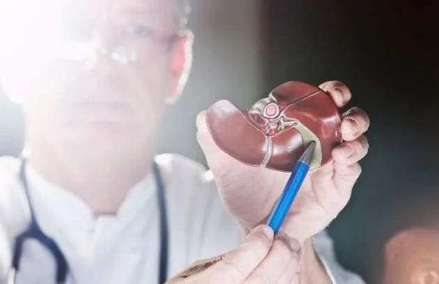
It is preferred for persistent tumors located close to the skin surface, particularly in areas such as the face or neck where aesthetic concerns are high, because it offers a non-surgical option. Similarly, it may be considered when a mass reappears after previous radiotherapy and the tissue can no longer tolerate another course of radiation treatment. It can also be applied to tumors located in deep internal organs such as the liver.
The main conditions for which this method is frequently considered are:
- Cutaneous tumors
- Malignant melanoma metastases
- Liver masses
- Head and neck recurrences
- Bone metastases
- Soft tissue sarcomas
- Vascular anomalies (Vascular malformations: Hemangioma, lymphangioma, AVM…)
How Does Reversible Electroporation Provide Benefits in Vascular Malformations?
Reversible electroporation technology is not limited to oncological treatments and has also begun to be used in the treatment of complex congenital vascular anomalies. These conditions, called vascular malformations, involve blood vessels developing abnormally and differently from normal, forming clusters. In cases where traditional vessel-sclerosing drug treatments (sclerotherapy) are insufficient, this new method may help increase the chances of successful treatment.
In this procedure, called electrosclerotherapy, a low dose of medication is administered into the abnormal vascular cluster, while electrical pulses are simultaneously delivered to the area under ultrasound guidance. The electrical current opens pores in the cells forming the abnormal vessel walls, making it easier for the medication to enter only these diseased cells. Thus, while the targeted vascular cluster gradually contracts and dries out, exposure of surrounding healthy tissues to the medication is largely prevented. Highly satisfactory cosmetic and functional results can be achieved.
What Are the Differences Between Reversible Electroporation and Other Heat-Based Methods?
Modern medicine offers many ablation (destruction) methods used to treat masses. Among these, systems based on thermal energy, such as radiofrequency ablation or microwave ablation, are quite common. These methods essentially aim to destroy cells by heating tissue to high temperatures, disrupting protein structures, and burning the cells. Although they are highly effective, their reliance on heat also brings certain physical disadvantages.
For example, if the mass being treated is located directly adjacent to a major blood vessel, the rapidly flowing blood inside the vessel acts as a coolant that carries heat away from the heated area. This is called the heat-sink effect in medicine. This situation may prevent the portion of the tumor near the vessel from heating sufficiently, allowing it to remain viable. Increasing the heat further, however, may damage the vessel wall. Reversible electroporation, on the other hand, uses a biophysical mechanism that is fundamentally not based on temperature changes. No significant heating or freezing occurs in the tissue during the procedure. Therefore, it may allow the lesion to be treated without damaging the vessel wall, independently of the cooling effect of blood flow.
How Does Reversible Electroporation Protect Healthy Tissues?
One of the issues patients are most curious about when masses are treated is the extent to which surrounding healthy tissues will be affected. Living cells are naturally sensitive to electrical current, and pores open in their membranes at certain voltages. However, the structures that hold the human body and organs together do not consist solely of cells. There are structural proteins and connective tissues that provide elasticity to blood vessels, surround nerves like a sheath, and give organs their shape.
This treatment method is designed to act largely on cell membranes alone. When electrical current is applied, the cell membranes open and the medication enters, but the supporting framework that holds these cells together is generally protected from serious damage caused by electrical stimulation. We can compare this to removing the furniture from a house while leaving its main load-bearing columns intact. Because the structural framework remains in place, the body’s own repair cells can migrate to this area over time and initiate the healing process.
The main structures that generally maintain their integrity during treatment are:
- Collagen fibers
- Elastin tissue
- Blood vessel walls
- Nerve sheaths
- Bile duct walls
Why Does Reversible Electroporation Not Produce the Known Side Effects of Chemotherapy?
In traditional systemic chemotherapy, drugs are administered through a blood vessel and circulate throughout the entire body. Although their purpose is to find and destroy cancer cells, they also tend to affect healthy cells that multiply rapidly in the body. This is why many patients may experience issues such as hair loss, extreme fatigue, or stomach problems. There are fundamental reasons why reversible electroporation differs in terms of these side effects.
First, the dose of medication used in this procedure is considerably lower than the amounts used in standard chemotherapy sessions. The second and most important reason is that the medication has a structure that prevents it from easily entering healthy cells even if it circulates freely throughout the body. The medication can enter only through the pores of cells in the area where the physician applies electrical current. Therefore, even if it reaches healthy organs, its potential to cause damage is very low because it cannot cross their cell membranes.
As a result, the main side effects that become less likely to occur in patients are:
- Hair loss
- Severe nausea
- Immune suppression
- Low platelet count
What Awaits Patients During the Recovery Process After Reversible Electroporation?
The fact that this treatment is performed using a minimally invasive approach, mostly through needle insertions in the skin, makes the subsequent recovery process significantly more comfortable compared with traditional surgeries. Because no large surgical incisions are made during the procedure, the need to wait for extensive wounds to heal or remain hospitalized for a long time is largely eliminated. Most patients are kept under observation for only a few hours until the effects of the anesthesia wear off.
Discomfort that may occur after the procedure generally consists of mild tenderness at the needle insertion sites or localized pain that can be managed with prescribed painkillers. Because the anatomical structure of the organs is preserved, patients can usually be discharged on the same day or the following day and return to their daily lives. Another advantage of this method is that it does not create permanent scar tissue, allowing it to be applied repeatedly to the same area if necessary.
The main factors supporting the recovery process are:
- No surgical incisions
- Short hospital stay
- Possibility of local anesthesia
- No scar formation in tissues
Why Should an Interventional Radiology Specialist Be Consulted for Reversible Electroporation?
Successfully planning and performing this treatment method on a patient requires much more than operating the device; it demands extensive anatomical knowledge and mastery of imaging technologies. Reaching a tumor hidden deep within the body and surrounded by vital blood vessels or nerve networks without surgery is a primary area of expertise for interventional radiology specialists who have focused specifically on this field during their medical training.
Interventional radiologists use radiological devices to read the inside of the human body like a transparent map, navigating through that map with needles at millimetric precision. For patients who have previously undergone numerous treatments, become exhausted by standard methods, or are considered at high risk for surgery, evaluation by these specialists may open new possibilities. Multidisciplinary evaluation of the patient’s condition by oncology boards and consideration of these minimally invasive treatment alternatives are extremely valuable steps in improving patient comfort and treatment adherence.
The main responsibilities of interventional radiology specialists during the process are:
- Image-guided needle placement
- Mapping of high-risk anatomical regions
- Minimally invasive procedure planning
- Post-procedure radiological follow-up evaluation





Vaka Örnekleri