


Medical oncology is the branch of medicine that plans and manages the drug-based treatment processes of cancer diseases after diagnosis. This field aims to control the tumor and prevent the spread of the disease by using systemic treatment methods such as chemotherapy, immunotherapy, targeted therapy, and hormone therapy.
The question of which diseases medical oncology deals with refers to a broad field that includes all types of cancer developing in different organs. Many malignant diseases such as breast cancer, lung cancer, colon cancer, stomach cancer, pancreatic cancer, and prostate cancer are among the main conditions followed and treated by medical oncology specialists.
The role of medical oncology in cancer treatment is to create a personalized treatment plan according to the stage and biological characteristics of the disease. Oncology specialists work in a multidisciplinary manner together with pathology, radiology, and surgical branches to determine the most appropriate drug treatment for the patient and regularly monitor the treatment process.
Medical oncology treatment methods are based on advanced drug technologies in modern cancer treatment. In addition to chemotherapy, smart drugs and immunotherapies aim to target cancer cells while preserving healthy tissues and contribute to increasing patients’ survival time and quality of life.
| Things You Should Know | Information |
| Definition of Medical Oncology | Medical oncology is the branch of medicine that includes the treatment of cancer with drugs. These treatments include chemotherapy, immunotherapy, targeted therapy (smart drugs), and hormone therapy. Medical oncologists plan the treatment after diagnosis, monitor the treatment process, and follow the course of cancer. |
| Role of the Oncologist | The oncologist plans and applies the treatment of patients diagnosed with cancer. They work in cooperation with other treatment methods such as surgery or radiotherapy and adopt a multidisciplinary approach. They also aim to increase patients’ quality of life and manage side effects. |
| Sub-branches of Oncology | In addition to medical oncology, there are two other main sub-branches: radiation oncology (radiation therapy) and surgical oncology (surgical removal of cancerous tissue). These three disciplines work together to carry out cancer treatment in a holistic manner. |
| Diseases Treated by Oncology | Oncology deals with malignant (cancerous) tumors that can develop anywhere in the body. These diseases include breast cancer, lung cancer, colorectal cancer, stomach cancer, prostate cancer, pancreatic cancer, lymphoma, leukemia, brain tumors, skin cancers (such as melanoma), and many other solid or hematological cancers. |
| Medical Oncology Follow-ups | During the treatment process, tumor responses are monitored with imaging and laboratory tests. In post-treatment periods, regular check-ups are also performed for disease recurrence. In addition, factors such as patients’ general condition, nutrition, psychological support, and palliative care are also monitored. |
| Cancer Treatment Methods | The main methods used in medical oncology are: Chemotherapy (drugs that kill cancer cells), Immunotherapy (treatment that helps the immune system recognize and target cancer cells), targeted therapy (drugs that attack biological targets specific to cancer cells), and hormone therapy (blocking hormone production in hormone-sensitive cancers). |
| Treatment Planning | Treatment is planned individually according to the patient’s general health condition, the type and stage of cancer, molecular characteristics, and the patient’s preferences. Treatment protocols are determined in accordance with national and international guidelines. |
| Multidisciplinary Approach | Medical oncologists usually work together with specialists from different branches such as surgeons, radiation oncologists, pathologists, radiologists, and when necessary, psychologists and nutritionists to determine the most appropriate treatment process for the patient. |
| Importance of Early Diagnosis | Early diagnosis in cancer significantly increases treatment success. Therefore, screening programs (mammography, colonoscopy, smear tests, etc.) are of great importance in individuals in the risk group. |
| Medical Oncology and Follow-up Process | After treatment, patients are called for follow-up at medical oncology outpatient clinics at certain intervals. During this process, it is evaluated whether the disease has recurred, and additional treatments are planned if necessary. |
What Is Medical Oncology and Which Systemic Treatments Does It Include?
Medical oncology is the branch that plans the drug-based treatment processes of individuals diagnosed with cancer from start to finish and develops personalized strategies according to the stage of the disease, its molecular structure, and the patient’s overall health status. Cancer cells have an incredible ability to adapt; they are very skilled at hiding from the immune system’s radar, creating new capillaries to feed themselves, and invading surrounding tissues. Medical oncology uses drugs from different classes and mechanisms of action precisely to break these harmful mechanisms and silence the signals that drive cell proliferation.
In the past, the most common and powerful weapon we had was chemotherapy drugs, which target the rate of cell division and destroy cells, but in doing so can also affect other rapidly dividing normal and healthy cells in our body such as hair follicles, the stomach lining, or bone marrow. Today, this situation has changed dramatically. By mapping the genetic and cellular structure of the tumor, smart drugs that hit only the weak point of the cancer cell without harming healthy tissues have come into use. Beyond that, instead of administering an external chemical substance, immunotherapy methods that train, stimulate, and enable the body’s own dormant immune system to attack cancer have opened a brand-new chapter in medical history. These treatments are sometimes used before surgery to shrink a large mass and make surgery possible, and sometimes after a successful surgery to eliminate invisible microscopic cancer cells that may be circulating in the blood and prevent the disease from returning.
The main systemic treatment methods applied in medical oncology clinics according to the structure of the disease are as follows:
- Chemotherapy
- Immunotherapy
- Smart drugs
- Hormone therapy
- Targeted agents
Which Diseases Does Medical Oncology Treat and What Types of Cancer Does It Focus On?
The possibility of cancer occurring in almost every organ and tissue of the body, from hair to nails, expands the field of oncology to an extremely broad dimension. This branch of science deals both with primary tumors that continue to grow in the tissue where they originate and with advanced-stage masses that have broken away and spread through the bloodstream to entirely different organs such as the lungs, liver, or bones. The incidence of cancers in society varies greatly depending on factors such as an aging population, changing dietary habits, environmental pollution, smoking, genetic heritage, and gender.
When it comes to human anatomy, very different disease groups emerge according to organ systems. For example, digestive system cancers include a line extending from the esophagus to the stomach and then to the small and large intestines, along with vital organs accompanying this line such as the pancreas, gallbladder, and liver. Urological cancers follow malignant growths in the kidney tissue, urinary tract, and bladder region. While cancers targeting female reproductive organs such as the uterus, ovaries, and cervix require highly specific approaches, diseases such as leukemia and lymphoma, which develop in the fluid nature of blood cells and lymph nodes, require entirely different treatment protocols. Sarcomas originating from bones and muscles in the skeletal system or tumors developing within brain tissue are also among the main targets of oncology.
The main cancer types commonly encountered in Türkiye and around the world and forming the daily practice of oncology are as follows:
- Lung cancer
- Breast cancer
- Prostate cancer
- Colorectal cancer
- Stomach cancer
- Pancreatic cancer
- Thyroid cancer
- Bladder cancer
- Cervical cancer
- Skin cancer
Which Biopsy Methods Does Interventional Radiology Use in Cancer Diagnosis?
For cancer to be treated accurately and effectively, it is never enough to know whether a lesion detected in imaging is cancerous. It is essential to determine exactly which cell type the mass originates from, its growth potential, the hormone receptors on its surface, and which drugs it resists or responds to. This is only possible by taking a sample from the mass and examining it under a microscope. In the past, obtaining samples from masses in internal organs required surgical incisions and anesthesia, but today modern imaging systems have fundamentally changed this process.
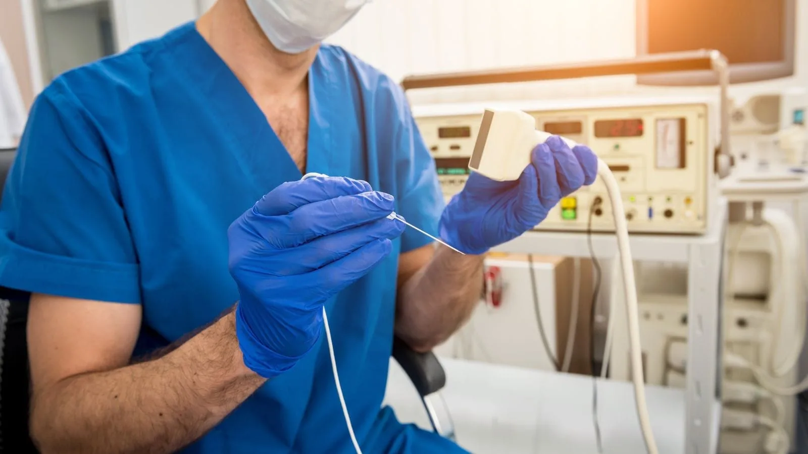
Interventional radiology is the strongest light that oncology shines into the darkness during the diagnostic phase. With special needles monitored in real time on the screens of high-resolution ultrasound devices, computed tomography scans that take millimetric slices, or magnetic resonance devices, the exact center of the suspicious tissue is safely reached. By making a tiny opening in the skin just large enough for a needle to pass through, the target is reached without damaging surrounding healthy tissues, major vessels, organ membranes, or nerve networks. These small tissue samples are sent to pathology laboratories and examined with special chemical stains. From the patient’s perspective, the greatest comfort is that these procedures are performed under local anesthesia, meaning only the needle entry site is numbed, the patient remains awake during the procedure, and they can return home on foot the same day after a few hours of observation.
Different medical needles are preferred during biopsy depending on the structure of the target organ. While very fine needles are used to aspirate cells from cystic fluid-filled structures or superficial lymph nodes under the skin, thicker spring-loaded and specially mechanized needles that preserve the tissue architecture are used for organs such as the liver, breast, prostate, or lungs.
The main imaging-guided biopsy procedures performed to confirm cancer diagnosis are as follows:
- Fine needle aspiration biopsy
- Core needle biopsy
- Core biopsy
- Vacuum-assisted biopsy
- Bone marrow biopsy
Why Is Port Catheter Use Important for Medical Oncology in Chemotherapy Treatment?
The route through which life-saving but equally potent chemotherapy drugs prescribed by medical oncology enter the body is a major detail that directly affects the comfort and continuity of the treatment process. These drugs contain highly concentrated chemicals by nature. During treatments lasting weeks or months, the thin superficial veins in our arms or on the back of our hands struggle to tolerate this irritating load. Over time, vein walls harden, veins become difficult to locate, blockages due to clotting occur, and most dangerously, there is a risk of the drug leaking out of the vein and causing burns under the skin. Trying to find a new vein with a needle at each treatment session becomes both a source of physical pain and a serious psychological barrier for the patient.
To completely eliminate this problem and secure the treatment line, special port systems are placed under the skin with a minimal intervention performed through a needle puncture. Usually placed a few centimeters below the right collarbone, a small reservoir is inserted that does not have any external tubes hanging, appearing only as a slight swelling under the skin. The flexible, very thin silicone tube attached to the end of this reservoir is directed into a large central vein in the neck region leading toward the heart, where liters of blood flow every second. As soon as the drug is administered through the port reservoir, it mixes into this large bloodstream and disperses diluted throughout the body within seconds without irritating any vein wall.
When patients come to the chemotherapy center, their arms are not repeatedly punctured with needles; the procedure begins by inserting a single special needle into this subcutaneous reservoir. These devices, which are placed in a completely sterile operating room or angiography environment under local anesthesia in a very short time and carry a very low risk of infection, do not restrict the patient’s daily life at all after the healing process is completed. The patient can bathe, swim, exercise, and use their arm comfortably.
The medical tools used to ensure safe vascular access in long-term oncological treatments are as follows:
- Port catheter
- Central venous catheter
- Tunneled catheter
- Temporary dialysis catheter
- Peripherally inserted central catheter
What Is Tumor Ablation and Which Diseases Does Non-Surgical Cancer Treatment Address?
In oncology practice, not every patient may have the physiological or anatomical conditions suitable for a standard major surgical operation. Advanced age, low cardiac and respiratory capacity, or the mass being located in a position attached to major vessels in the liver that makes it inoperable can render surgical intervention impossible. Sometimes, for a few small tumors that have spread to other organs, it is not desirable to subject the patient to the stress of major general anesthesia and surgery. At such critical turning points, ablative treatments—the most refined and technological field of interventional oncology—come into play, involving the destruction of the mass in place by burning or freezing it.
In its simplest form, the concept of ablation means that instead of cutting out the cancerous tissue from an organ, special needles are used to reach the exact center of that tissue and destroy it in place using different physical energies. The target tissue is clearly mapped in three dimensions on a computed tomography screen. After numbing the skin, special antennas that can open like an umbrella tip are placed into the core of the tumor. If heat energy is preferred, high-frequency radio waves or microwave energy emitted from the tip of the needle rapidly vibrate the water molecules inside the mass, generating heat up to 100 degrees within seconds. The tumor essentially burns without flames and turns into completely dead tissue. If freezing is used, high-pressure special gases are delivered into the needle, cooling its tip down to minus 80 degrees and turning the cancerous tissue into a large ice ball inside the body.
There are also electricity-based ablation systems developed for tumors located in more specific areas. In this system, no heat energy is produced; instead, extremely high-voltage electrical shocks are delivered through the needles for a very short duration. These shocks create irreparable pores in the membranes of cancer cells, causing the tumor to dissolve, while not causing any permanent physical damage to major blood vessels, bile ducts, or urinary pathways adjacent to the tumor. After these procedures, which do not require general anesthesia and keep pain at a minimum level, the patient can continue their life the next morning after staying in the hospital for just one night.
The ablation methods used in interventional oncology to destroy cancer tissue in place without surgery are as follows:
- Radiofrequency ablation
- Microwave ablation
- Cryoablation
- Electroporation
- Laser ablation
- Alcohol ablation
What Are Intravascular Treatments Such as TACE and TARE in Liver Cancers?
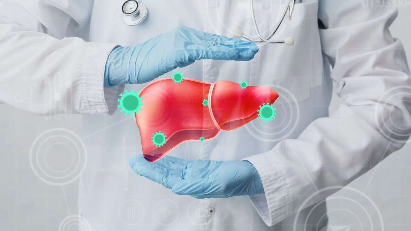
The liver is a massive, sponge-like, and incredibly active chemical purification plant in our body. All blood flow coming from the stomach, intestines, and pancreas is directly filtered through the liver. Unfortunately, this unique vascular network also turns the liver into a perfect landing and lodging site for cancer cells that travel through the bloodstream from organs such as the colon, breast, stomach, or neuroendocrine glands. In addition, there are primary carcinomas that arise from the liver’s own healthy cells undergoing transformation.
While fighting all these liver cancers, we have a remarkable anatomical advantage provided by nature. In the miraculous structure of the liver, healthy tissue receives almost all of its nutrients and oxygen from a massive vein called the portal vein, which filters blood coming from the intestines. However, the cancer cells that settle in the liver are extremely greedy; to grow and multiply rapidly, they latch onto the arterial system called the hepatic artery, which carries fresh oxygen-rich blood. Interventional oncology turns this vulnerability of the tumor to its advantage. Under local anesthesia, a journey is made through the vessels to the liver using very thin, spaghetti-like catheters inserted through the artery in the groin or wrist, and the oxygen-rich vessel directly feeding the tumor is identified.
If the goal is to deliver intensive medication and block the vessel, the “TACE” method is applied. From the tip of the catheter, microscopically sized sponge-like spheres containing oncology drugs are injected into the tumor vessel. These spheres act as plugs, blocking the tumor’s feeding vessel within seconds and suffocating the cancer, while simultaneously releasing the concentrated drug directly into the tumor for days without harming surrounding tissues. If the lesion is very large or has infiltrated vascular beds, the “TARE” method, or radioembolization, is preferred. This time, instead of drugs, the spheres are loaded with a very special nuclear isotope called Yttrium-90, which affects only a few millimeters around it. These radioactive atoms injected into the tumor bed irradiate the mass intensely from the inside out, destroying it as if with a microscopic nuclear attack.
The artery-based interventional treatment methods used to target tumors within the liver are as follows:
- Transarterial chemoembolization
- Transarterial radioembolization
- Hepatic arterial infusion
- Direct intra-arterial chemotherapy
- Bland embolization
What Interventions Improve Quality of Life in Advanced-Stage Oncology Patients?
Oncology practice is not only concerned with eliminating the disease from the body or prolonging life; one of its most sacred duties is to alleviate the destructive effects of the disease on the body and ensure that every remaining day of the patient is lived under humane conditions, without pain, in peace and comfort. As tumors grow uncontrollably within the body, they form masses that occupy natural spaces and exert serious mechanical pressure on surrounding delicate structures, fluid channels, or nerve bundles. The blockages that occur as a result of this pressure or the disruption of internal fluid balances of organs can quickly endanger the patient’s vital functions.
For example, a pancreatic mass pressing on the bile ducts at the liver outlet can cause bile to mix into the blood, leading to complete yellowing of the skin, severe itching that prevents sleep, nausea, and life-threatening infections due to bacteria entering the bloodstream. Similarly, when ovarian or bladder tumors obstruct the channels through which urine flows from the kidneys to the bladder, urine backs up into the kidney and can lead to organ failure within just a few days. When tumors spread to organ membranes, liters of fluid can accumulate in the abdominal cavity or in the chest cavity around the lungs. This massive fluid accumulation in the abdomen puts pressure on the diaphragm, making it impossible for the patient to take deep breaths or even eat a few bites, while creating an unbearable sense of tension.
At these stages, when major and exhausting surgical operations are not an option, high-tech palliative interventions provide relief. Under ultrasound and fluoroscopy guidance, flexible special metal or plastic mesh tubes (stents) are placed into the obstructed bile ducts or urinary pathways through tiny needle punctures in the skin, restoring normal fluid flow within hours. These compressive fluids accumulated in the lungs or abdomen are drained outside the body with very thin soft tubes, providing immediate relief. In cases of severe nerve-related pain where even painkillers are insufficient, the nerve networks behind the abdomen are reached and numbed with alcohol, breaking the cycle of intense suffering.
The palliative procedures applied to solve mechanical problems caused by advanced-stage cancers and provide comfort are as follows:
- Biliary stenting
- Biliary drainage catheter placement
- Percutaneous nephrostomy
- Ureteral stenting
- Pleural fluid drainage
- Ascites fluid drainage
- Celiac ganglion block
- Tumor bleeding embolization
What Is a Multidisciplinary Tumor Board in Cancer Treatment and Why Is It Vital?
Cancer is an equation with many variables in which the organism rewrites its own rules. Thanks to the dizzying medical advances of the modern era, no specialist physician can claim to make the most accurate and flawless decision by reviewing a patient’s file alone behind closed doors. To bring the disease under control, every branch of medicine must put its own arsenal and perspective on the table. Tumor boards, which are the central decision-making mechanism of modern medical oncology practice, serve this purpose as collective intelligence summits where physicians from different specialties come together to determine the best path for the patient.
In these meetings, there is a team around a large table examining every detail. When the meeting begins, the pathologist explains the rebellious nature of the tumor under the microscope and the genetic codes of the cells. Based on this information, the medical oncologist determines which smart drug, chemotherapy agent, or immunotherapy method will systemically weaken the tumor. Then the surgeon evaluates anatomically whether the mass is attached to major vessels and whether the patient can tolerate the stress of such an operation. The radiation oncologist outlines the limits of how much surrounding organs may be affected if external radiation is applied. And right in the middle of this critical discussion, the interventional radiologist takes the floor and, using 3D tomography images projected onto large screens, demonstrates how they can safely reach that suspicious, otherwise inaccessible lesion with a needle for biopsy or how they can enter through the vessels and eliminate the mass without any incision in a patient for whom surgery is risky.
This interdisciplinary brainstorming dramatically accelerates the treatment process by saving the patient from moving between departments and unnecessary delays. It offers not the most aggressive treatment, but the most rational, targeted, and least harmful treatment combination appropriate to the patient’s current physiological condition. These consensus decisions maximize the patient’s chance of recovery in light of scientific literature.
The main medical disciplines that collaboratively draw the patient’s treatment roadmap in tumor boards are as follows:
- Medical oncology
- Surgical oncology
- Radiation oncology
- Interventional radiology
- Pathology
- Nuclear medicine
Frequently Asked Questions
What Is Medical Oncology and what role does it play in cancer care?
Medical oncology is a medical specialty focused on the diagnosis, treatment, and management of cancer. Medical oncologists develop personalized treatment plans and coordinate care with surgeons, radiation oncologists, and other specialists.
Which Diseases Does Oncology Treat besides common cancers?
Oncology primarily treats cancers affecting various organs and tissues, including breast, lung, colon, prostate, blood, and skin cancers. It also manages certain precancerous conditions and cancer-related complications.
How does a medical oncologist diagnose and evaluate cancer?
Medical oncologists use medical history, physical examinations, laboratory tests, imaging studies, biopsies, and molecular testing to determine the type, stage, and characteristics of a patient’s cancer.
What treatment methods are commonly used in medical oncology?
Medical oncology treatments may include chemotherapy, immunotherapy, targeted therapy, hormone therapy, and other systemic treatments designed to destroy cancer cells or slow their growth throughout the body.
Can Medical Oncology be combined with surgery or radiation therapy?
Yes, medical oncology is often part of a multidisciplinary approach. Systemic treatments may be given before or after surgery or combined with radiation therapy to improve treatment effectiveness and outcomes.
How does personalized medicine influence oncology treatment plans?
Advances in genetic and molecular testing allow oncologists to tailor treatments based on specific tumor characteristics. Personalized therapies may improve effectiveness while reducing unnecessary side effects for some patients.
What side effects can occur during oncology treatments?
Side effects vary depending on the treatment used and may include fatigue, nausea, hair loss, appetite changes, immune suppression, or skin reactions. Many side effects can be managed with supportive care.
Who may benefit from regular oncology screenings and evaluations?
Individuals with a family history of cancer, genetic risk factors, previous cancer diagnoses, or certain lifestyle-related risks may benefit from screening programs and regular oncology evaluations.
How often are patients monitored during medical oncology treatment?
Monitoring schedules depend on the cancer type and treatment plan. Patients typically undergo regular examinations, blood tests, and imaging studies to assess treatment response and detect potential complications.
Can early diagnosis improve outcomes in oncology patients?
Early cancer detection often allows treatment at a less advanced stage, increasing the likelihood of successful therapy, reducing complications, and improving long-term survival and quality of life.

Prof. Dr. Özgür Kılıçkesmez is one of Türkiye’s leading interventional radiology and neuroradiology specialists, focusing on minimally invasive endovascular treatments. His primary areas of expertise include aneurysm and stroke treatments, embolization procedures, non-surgical treatments of thyroid and parathyroid diseases, interventional oncology, and peripheral vascular interventions. He currently serves as the Head of the Interventional Radiology and Neuroradiology Department at Istanbul Memorial Göztepe Hospital.
Graduating from Istanbul University Cerrahpaşa Faculty of Medicine in 1997, Prof. Dr. Kılıçkesmez received advanced training in interventional radiology and oncology as a Clinical Fellow at London Guy’s & St Thomas’ and King’s College Hospital between 2012 and 2013. He earned the title of Professor in 2020 and, in the same year, became the Founding Interventional Radiologist at SBÜ Çam and Sakura City Hospital. He also holds the EBIR (European Board of Interventional Radiology) certification.
Prof. Dr. Kılıçkesmez has 94 international and 40 national publications and over 2500 citations. He serves as a reviewer for prestigious journals such as the American Journal of Roentgenology (AJR) and Diagnostic & Interventional Radiology. With his clinical and academic expertise, he is a nationally and internationally recognized authority in stroke treatment, brain aneurysm, embolization, tumor ablation, and minimally invasive treatments of thyroid/parathyroid diseases.





Vaka Örnekleri