


Jugular vein compression is the condition in which the internal jugular vein, which carries metabolic waste and deoxygenated blood from the brain back to the heart, is mechanically compressed by the styloid process, the C1 vertebra, or muscular structures in the neck region, thereby obstructing blood flow. This venous narrowing disrupts the absorption of cerebrospinal fluid and increases intracranial pressure; it leads to neurological symptoms that reduce quality of life such as persistent headaches, pulsatile tinnitus (hearing the heartbeat in the ear), visual disturbances, and brain fog. This condition, also known in the medical literature as Styloidogenic Jugular Venous Compression Syndrome (SJVCS), is not merely a simple neck pain but a serious vascular pathology that disrupts cerebral circulatory balance and requires hemodynamic evaluation.
What Is Jugular Vein Compression and Why Is It Important?
We can liken our circulatory system to a city’s water network. Just as there are pipes that bring clean water, there are also drainage pipes that remove used water. In our body, we call the vessels that carry clean blood arteries, and those that collect deoxygenated blood veins. The internal jugular vein is the “main drainage pipe” of our brain. The brain, as the most energy-consuming organ in the body, works continuously and produces a large amount of waste. These wastes and deoxygenated blood must be safely removed from the skull and returned to the heart.
In a healthy individual, this flow is silent and uninterrupted. However, this main vessel passes through a narrow tunnel in the neck region. If there is a “traffic accident” or narrowing in this tunnel, meaning the vessel is subjected to external pressure, the drainage of blood becomes difficult. Just as water backs up when a sink pipe is clogged, deoxygenated blood in the brain accumulates backward. This increases intracranial pressure and disrupts the brain’s working environment. For us physicians, this is not merely an anatomical narrowing but a serious physiological problem that disrupts the patient’s overall comfort.
What Causes Jugular Vein Compression?
Many patients struggle to understand why they experience such a problem without having had any accident. The root of the problem is usually that the bone and soft tissue structures in the neck do not “leave space” for the vein. After exiting the skull, the jugular vein descends between the cervical vertebrae and certain bony prominences. During this journey, its biggest adversary is usually the bone we call the “styloid process.”
This bony projection, which extends downward just below the ear, is congenitally longer in some individuals or has a different angle. This elongated bone compresses the jugular vein passing right next to it between itself and the posterior cervical vertebra (C1). Just like a nutcracker crushing a walnut, the vein caught between the two bones is compressed. This is called “Styloidogenic Jugular Venous Compression Syndrome” (SJVCS).
In addition, there may be other factors that compress the vein:
- Hypertrophy of neck muscles
- Swelling of lymph nodes
- Structural tortuosity of the carotid artery passing nearby
- Calcified connective tissues
What Are the Symptoms of Jugular Vein Compression?
The symptoms of this syndrome can vary greatly from person to person and usually progress “insidiously.” Patients are often treated for years with diagnoses such as “migraine” or “psychogenic pain,” yet their complaints do not resolve, because the problem is vascular. The most characteristic feature is that symptoms change with head position.
The most commonly reported complaints by our patients are:
- Severe headaches, especially in the morning
- A feeling of fullness and pressure in the head
- Pain that increases with coughing or straining
- Pain radiating from the neck to the eye
- Blurred vision or temporary visual dimming
- A sensation of pressure behind the eyes
- Chronic fatigue
- Sleep disturbances
- Restricted neck movements
What Is Pulsatile Tinnitus?
One of the most prominent and distressing reasons our patients visit the clinic is “pulsatile tinnitus.” Typical tinnitus is usually a high-pitched “ringing” sound. However, those with pulsatile tinnitus hear their own heartbeat in their ears. This sound is rhythmic and synchronized with the pulse.
Patients describe this sound as:
- A whooshing wind-like noise
- The sound of flowing water
- A rhythmic “whoosh-whoosh” sound
- A pounding heartbeat in the ear
This sound is not imaginary. When the vessel is compressed, the blood passing through it becomes turbulent. Just like when you squeeze the end of a garden hose and water spurts out with a sound, blood passing through a narrowed vessel also produces noise. Since the vessel is very close to the ear, the patient can hear this sound. The fact that the sound changes when slight pressure is applied to the neck or when the head is turned is the strongest evidence that the problem is vascular.
Why Does Brain Fog Occur?
Beyond physical pain, one of the most challenging conditions for our patients is mental cloudiness. Slowing of blood circulation in the brain and the inability to clear waste products (metabolites) quickly enough reduce the efficiency of brain cells.
The effects caused by this condition include:
- Forgetfulness
- Difficulty finding words
- Difficulty concentrating
- Constant feeling of sleepiness
- Mental fatigue
- Focus problems
Our patients describe this as “there is a cloud inside my head” or “I feel like I am watching the world from behind a glass dome.” When venous flow improves after treatment, patients saying “my mind has cleared” clearly demonstrates the cognitive effects of circulatory impairment.
How Is It Diagnosed?
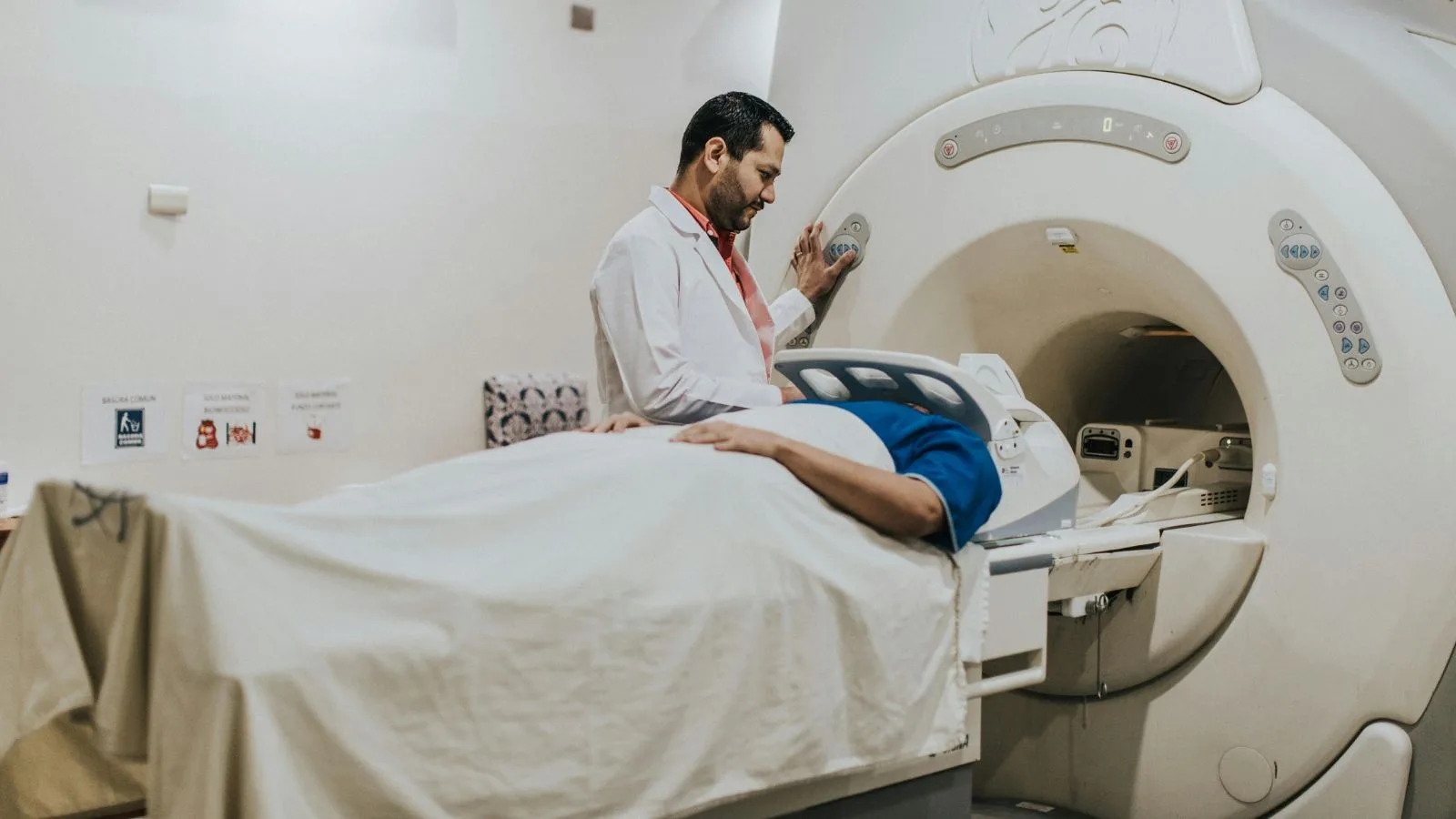
The diagnostic process begins with careful listening to the patient, but advanced imaging methods are essential for a definitive diagnosis. A standard neck ultrasound or X-ray may be insufficient to show this deep compression behind the bones. As interventional radiologists, we approach the situation more technically and in three dimensions.
The first step is usually Computed Tomography Venography (CTV). In this procedure, contrast material is administered through a vein and the neck region is imaged. However, there is a very critical detail here: dynamic imaging. Images should be taken not only while the patient is lying flat but also when the head is turned to the right and left. Because sometimes the vein may be open in a neutral position but can become completely occluded when the head is turned and gets trapped between the bony structures.
Other methods we use in diagnosis include:
- MR Venography (MRV)
- Dynamic Doppler Ultrasonography
- Three-dimensional reconstructions
- Detailed neck tomography
What Is Venous Manometry and Why Is It Necessary?
Imaging methods show us the anatomical structure, that is, how much the vessel is narrowed. However, they do not answer the question: “Is this narrowing truly the cause of the patient’s symptoms?” A vessel that appears very narrow on imaging may sometimes cause no symptoms because the body has developed collateral pathways. This is where “venous manometry” comes into play.
Manometry is a pressure measurement procedure performed in an angiography suite. A very thin catheter is inserted through the groin and advanced to the jugular vein. The blood pressure before and after the narrowing is measured. If the pressure behind the narrowing is significantly higher and drops after passing it, this indicates a “hemodynamically significant obstruction.” We also perform this measurement while turning the head to the right and left. If the pressure difference (gradient) is high, it scientifically proves that the patient’s headache and noise are caused by this narrowing. This is the most important test guiding the treatment decision.
How Is Jugular Vein Compression Treated?
The treatment plan is individualized based on the cause and severity of the narrowing. Not every patient requires immediate intervention; in mild cases, follow-up and lifestyle modifications may be sufficient. However, in patients with severe symptoms and high pressure confirmed by manometry, more radical solutions are required. The main goal of treatment is to relieve the pressure on the vessel and restore normal blood flow.
Treatment options include:
- Medical therapy
- Neck exercises
- Surgical decompression
- Balloon angioplasty
- Stenting
- Combined (hybrid) treatments
What Is Surgical Decompression?
If the main cause of the narrowing is the length of the bone called the “styloid process,” the most logical and permanent solution is shortening this bone. This is called “styloidectomy.” In this surgery, performed by ENT or neurosurgery specialists, the bone segment compressing the vein is removed.
The advantage of this method is that it resolves the problem at its root without placing a foreign body (stent) inside the vessel. Once the bone pressure is removed, the vessel usually relaxes and regains its original form. However, in some cases, the vessel may have lost its elasticity due to prolonged compression and may not fully expand even after the bone is removed. In such cases, additional treatments may be required.
When Is Stent Treatment Applied?
As interventional radiologists, we frequently use stents (metal mesh) in vascular narrowings. However, the use of stents in jugular vein compression is a delicate matter. If a rigid bone is compressing the vessel from the outside, placing a stent there can be risky. The strong bone structure can crush, fracture, or deform the metal stent, leading to more serious problems.
Therefore, we generally prefer stent treatment in the following situations:
- Narrowings without bony compression
- Cases where the vessel does not open sufficiently after surgery
- Intraluminal membranes or adhesions
- Acute thrombosis cases
The ideal approach is usually a “hybrid” method. First, the bony compression is removed surgically. After the healing process, if the vessel is still narrow, a stent can be safely placed since there is no longer a risk from bone compression, restoring full patency of the vessel.
What Is the Recovery Process After Treatment?
When treatment is successful, the improvement in patients’ quality of life is quite dramatic. Symptoms related to venous pressure, in particular, regress rapidly. Even immediately after the procedure or surgery, patients may say “that heaviness in my head is gone.”
Observed improvements during recovery include:
- Resolution of ear noise
- Reduction in headaches
- Improved visual clarity
- Better sleep quality
- Increased mental energy
Full recovery may sometimes take several weeks or months. Especially the resolution of edema behind the eyes and the brain’s adaptation to the new circulatory pattern may take time. During this process, regular use of blood-thinning medications is very important.
Is There a Risk of Thrombosis?
Wherever blood flow slows down, the risk of clotting increases. Narrowing in the jugular vein can cause pooling of blood in the cerebral sinuses. This condition, although rare, may lead to “Cerebral Venous Sinus Thrombosis.” This is a serious condition and requires urgent intervention.
Symptoms of thrombosis include:
- Sudden and severe headache
- Seizures
- Numbness on one side of the body
- Confusion
- Severe vomiting
In such cases, as interventional radiologists, we enter via angiography and remove the clot (thrombectomy). Therefore, monitoring patients with jugular vein compression is vital in managing these risks.
The Importance of a Multidisciplinary Approach
Jugular vein compression is not a condition that can be managed by a single physician alone. Collaboration between different specialties is required from diagnosis to treatment. The neurologist manages the patient’s clinical follow-up, we (interventional radiologists) map the vessel and measure pressures, and surgeons remove the mechanical obstruction.
Thanks to this teamwork:
- Unnecessary procedures are avoided
- The most appropriate treatment plan is selected
- The risk of complications is reduced
- The patient is evaluated holistically
The most important thing our patients should know is that this challenging condition they experience is not without a solution. This anatomical reality underlying many complaints that are dismissed as “psychological” can be treated with the right methods in the right hands.
What Should Be Considered in Daily Life?
There are some simple precautions that diagnosed or suspected patients should pay attention to in daily life. These measures can help balance intracranial pressure.
Points to consider include:
- Sleeping with a high pillow
- Drinking plenty of water
- Avoiding movements that strain the neck
- Preventing constipation
- Reducing excessive salt intake
- Avoiding prolonged head-down positions
Frequently Asked Questions
Jugular Vein Compression affect blood drainage from the brain?
Jugular vein compression can restrict the normal outflow of blood from the brain, potentially increasing venous pressure. Reduced drainage may contribute to neurological symptoms and can affect overall cerebral circulation in some individuals.
Jugular Vein Compression cause headaches and pressure-related symptoms?
Jugular vein compression may contribute to chronic headaches, head pressure, dizziness, and visual disturbances. Symptoms can occur when impaired venous drainage affects intracranial pressure and normal blood flow dynamics.
Jugular Vein Compression associated with pulsatile tinnitus?
Jugular vein compression has been linked to pulsatile tinnitus in some patients. Altered blood flow patterns near the compressed vein can create abnormal vascular sounds that are perceived as rhythmic noises in the ear.
Jugular Vein Compression develop from anatomical abnormalities?
Jugular vein compression may result from elongated styloid processes, enlarged cervical vertebrae, muscular structures, or other anatomical variations. These structures can narrow the vein and interfere with normal venous outflow.
Jugular Vein Compression diagnosed through imaging studies?
Jugular vein compression is commonly evaluated using CT venography, MR venography, Doppler ultrasound, or catheter-based venography. These studies help identify the location, severity, and potential causes of venous narrowing.
Jugular Vein Compression increase the risk of intracranial hypertension?
Jugular vein compression may contribute to elevated intracranial venous pressure in certain patients. When cerebral venous drainage becomes impaired, symptoms associated with intracranial hypertension can sometimes develop or worsen.
Jugular Vein Compression treated without surgery?
Treatment options may include observation, symptom management, and addressing contributing medical conditions. Conservative approaches can be appropriate for mild cases, while more severe compression may require interventional evaluation.
Jugular Vein Compression require surgical treatment in severe cases?
Surgery may be considered when significant symptoms are clearly linked to anatomical compression and conservative measures are unsuccessful. The specific procedure depends on the underlying structure causing the venous obstruction.
Jugular Vein Compression recovery after treatment vary between patients?
Recovery timelines depend on symptom severity, treatment type, and overall health. Some individuals experience improvement within weeks, while others may require longer follow-up to assess changes in venous drainage and symptoms.
Jugular Vein Compression lead to long-term complications if untreated?
Persistent jugular vein compression may contribute to ongoing headaches, pulsatile tinnitus, visual symptoms, or quality-of-life concerns. Early evaluation can help determine whether treatment is needed to prevent symptom progression.

Prof. Dr. Özgür Kılıçkesmez is one of Türkiye’s leading interventional radiology and neuroradiology specialists, focusing on minimally invasive endovascular treatments. His primary areas of expertise include aneurysm and stroke treatments, embolization procedures, non-surgical treatments of thyroid and parathyroid diseases, interventional oncology, and peripheral vascular interventions. He currently serves as the Head of the Interventional Radiology and Neuroradiology Department at Istanbul Memorial Göztepe Hospital.
Graduating from Istanbul University Cerrahpaşa Faculty of Medicine in 1997, Prof. Dr. Kılıçkesmez received advanced training in interventional radiology and oncology as a Clinical Fellow at London Guy’s & St Thomas’ and King’s College Hospital between 2012 and 2013. He earned the title of Professor in 2020 and, in the same year, became the Founding Interventional Radiologist at SBÜ Çam and Sakura City Hospital. He also holds the EBIR (European Board of Interventional Radiology) certification.
Prof. Dr. Kılıçkesmez has 94 international and 40 national publications and over 2500 citations. He serves as a reviewer for prestigious journals such as the American Journal of Roentgenology (AJR) and Diagnostic & Interventional Radiology. With his clinical and academic expertise, he is a nationally and internationally recognized authority in stroke treatment, brain aneurysm, embolization, tumor ablation, and minimally invasive treatments of thyroid/parathyroid diseases.




Vaka Örnekleri