


Cerebral venous manometry is a specialized diagnostic method used to measure blood pressure in the brain’s vessels. This procedure provides critical information in the diagnosis and treatment of various neurological diseases by precisely measuring pressure changes in the venous system (veins) of the brain. It plays an important role especially in the evaluation of conditions such as intracranial hypertension (increased pressure inside the skull) and venous sinus thrombosis (clot formation in the brain veins).
In Which Conditions Is Cerebral Venous Manometry Applied?
Cerebral venous manometry is used in the diagnosis and treatment planning of various problems related to brain vessels. The most common conditions in which this procedure is applied are:
- Idiopathic intracranial hypertension (formerly known as pseudotumor cerebri)
- Venous sinus stenosis (narrowing in brain veins)
- Suspicion of venous sinus thrombosis
- Resistant headaches
- Presence of papilledema (swelling of the optic nerve)
- Pulsatile tinnitus (heartbeat-synchronous ringing in the ear)
- Visual disturbances and visual field loss
Especially in cases where a definitive diagnosis cannot be made with other imaging methods or when there is no response to treatment, cerebral venous manometry can provide valuable information. By directly measuring pressure gradients, this procedure can reveal functional problems that imaging methods such as MRI or CT cannot show.
What Is the Preparation Process Before the Procedure?
Before cerebral venous manometry, a comprehensive preparation process is required to ensure the procedure is performed safely and effectively. This process generally includes the following steps:
Medical Evaluation and Tests:
- Detailed neurological examination
- Blood tests (coagulation parameters, kidney functions)
- Imaging studies (MR venography, CT angiography)
- Fundoscopic examination (to assess the presence of papilledema)
Medication Adjustments:
- Blood thinners (aspirin, warfarin, DOACs) are usually discontinued 3-7 days before the procedure
- Special adjustments may be made for diabetes medications
- You must inform your doctor about any medications you are allergic to
Preparation on the Day of the Procedure:
- You will be asked to fast for 6-8 hours before the procedure
- Your doctor will guide you on taking your morning medications with a small amount of water
- It is recommended to wear comfortable clothing and leave valuables at home
- You should have a relative accompany you, as it will not be appropriate for you to drive after the procedure
Before the procedure, your doctor will provide detailed information and answer your questions. At this stage, expressing all your concerns and questions is important for the success of the procedure.
How Is the Cerebral Venous Manometry Procedure Performed?
Cerebral venous manometry is a minimally invasive procedure usually performed in an angiography unit under imaging guidance. The procedure consists of the following stages:
- Preparation and Monitoring:
- When you are taken to the procedure room, your heart rate, blood pressure, and oxygen level will be monitored by connecting you to monitors
- The groin area is cleaned and covered with sterile drapes to ensure a sterile environment
- Local Anesthesia and Vascular Access:
- Local anesthesia is usually applied to the femoral vein in the groin area
- Access is obtained with two catheters through both the femoral artery and the femoral vein.
- A small needle is used to enter the vessel, and then a guidewire is placed
- A thin catheter (tube) is inserted into the vessel over the guidewire
- Advancement of the Catheter:
- The catheter is advanced under fluoroscopy (real-time X-ray) guidance, starting from the femoral vein, passing through the right side of the heart, and reaching the brain’s venous sinuses via the neck vessels
- At this stage, contrast material may be administered to visualize the vessels
- Pressure Measurements:
- The catheter is positioned in different venous regions of the brain (superior sagittal sinus, transverse sinus, sigmoid sinus, jugular bulb) and pressure measurements are taken
- Pressure values are digitally recorded and analyzed
- If necessary, measurements can be repeated in different positions (lying, sitting)
- Therapeutic Procedures (If Necessary):
- In some cases, therapeutic interventions (such as venous stenting) may be performed in the same session following the diagnostic procedure
- Completion of the Procedure:
- After all measurements are completed, the catheter is removed
- Manual pressure is applied to the entry site to stop bleeding
- A sterile dressing is applied to the area
The procedure usually takes 1-2 hours, but this duration may be extended if additional interventions are performed. Throughout the entire process, the medical team performing the procedure will remain in communication with you and continuously evaluate your condition.
What Does the Patient Feel During the Procedure?
Cerebral venous manometry is usually performed under local anesthesia and is a well-tolerated procedure for most patients. The sensations you may experience during the procedure include:
- During Local Anesthesia: You may feel a brief burning or stinging sensation during the local anesthesia injection in the groin area. This sensation usually disappears within a few seconds.
- During Catheter Advancement: You generally do not feel anything as the catheter moves inside the vessel. Some patients may describe a mild pressure or pulling sensation, but it is not uncomfortable.
- During Contrast Injection: When contrast material is injected, you may feel a temporary warmth in your body, a metallic taste, or an urge to urinate. This is normal and usually resolves within 1-2 minutes.
- During Pressure Measurements: You will not feel pain while pressure measurements are taken. In some patients, especially those with high intracranial pressure, temporary headache may occur during catheter manipulation.
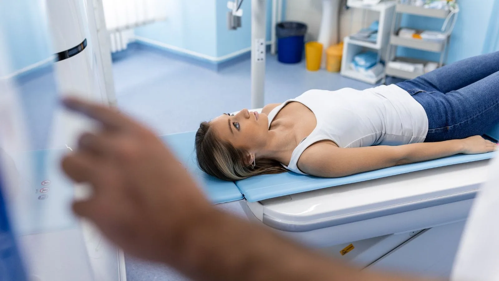
- Position During the Procedure: You will need to lie still on your back during the procedure. Staying in the same position for a long time may cause discomfort in some patients.
- In some cases, measurements may be repeated in different head positions, and separate measurements may be taken from the right and left sides. In this way, the effect of dynamic changes on pressure can be demonstrated.
If you feel any discomfort during the procedure, you must inform the medical team performing it. If necessary, additional sedation or pain relief may be administered. Throughout the procedure, the healthcare team will remain in communication with you and continuously monitor your condition.
What Are the Risks and Side Effects of Cerebral Venous Manometry?
Providing clear, honest, and transparent information about the risks, side effects, and limitations of the procedure is critical for patients to make informed decisions. As with any medical procedure, cerebral venous manometry has certain risks and may not be suitable for everyone.
Although cerebral venous manometry is generally a safe procedure, the following risks and side effects may occur:
Common Side Effects:
- Bruising or tenderness at the entry site
- Mild headache
- A feeling of fatigue that may last for a few hours after the procedure
- Nausea (usually related to the contrast material)
Less Common Complications:
- Bleeding or hematoma (blood accumulation) at the entry site
- Clot formation within the vessel (thrombosis)
- Allergic reactions to contrast material
- Temporary heart rhythm disturbances
Rare Serious Complications:
- Stroke or transient ischemic attack
- Vessel perforation
- Infection
- Impairment of kidney function (due to contrast material)
Most of these risks can be minimized with the experience of the medical team performing the procedure and the precautions taken. Before the procedure, your personal risk factors will be evaluated to determine the safest approach for you.
What Are the Limitations of the Procedure?
Although cerebral venous manometry is a valuable diagnostic tool, it also has some limitations:
- Technical Difficulties: In some patients, advancing the catheter may be difficult due to anatomical variations or previous vascular diseases.
- Measurement Variables: Pressure measurements may be affected by factors such as the patient’s position, hydration status, and depth of anesthesia.
- Interpretation Differences: There may be some differences between centers in interpreting measurement results.
- Contraindications: The procedure may not be performed or may be postponed in the following situations:
- Uncontrolled bleeding disorders
- Severe kidney failure
- Contrast material allergy
- Pregnancy (especially the first trimester)
- Presence of active infection
- Limited Availability: This specialized procedure may not be available in every center and may require referral to experienced centers.
These limitations require an individualized risk-benefit assessment for each patient. Your doctor will decide whether this procedure is appropriate for you based on your specific condition.
How Does the Post-Procedure Process Progress?
The recovery process after cerebral venous manometry is generally quick, and most patients can be discharged on the same day. The post-procedure process proceeds as follows:
First Hours (Post-Procedure Observation):
- You will be kept under observation for 2-6 hours after the procedure
- Your vital signs (blood pressure, pulse, respiration) will be regularly monitored
- The entry site will be checked for bleeding or swelling
- Your neurological status will be evaluated
Discharge and Return Home:
- You will be discharged once your condition is stable
- You should not drive on the day of the procedure; it is recommended that a relative takes you home
- It is important to keep the dressing at the entry site clean and dry
- Avoid heavy physical activities for 24-48 hours
First Days:
- Drinking plenty of fluids helps eliminate the contrast material from the body
- Mild pain relievers (such as paracetamol) are usually sufficient for discomfort at the entry site
- If there is increasing redness, swelling, pain, or fever at the entry site, you should consult your doctor immediately
Follow-Up Process:
- Procedure results are usually evaluated within 1-2 days
- Your treatment plan is determined or adjusted according to the results
- Your follow-up appointment is usually scheduled within 1-4 weeks
- In some cases, additional imaging studies or therapeutic interventions may be required
Carefully following the specific instructions given to you after the procedure will reduce the risk of complications and accelerate your recovery process.
Summary
Cerebral venous manometry is a valuable diagnostic method that plays a critical role in diagnosing neurovascular problems that cannot be detected by other diagnostic methods by directly measuring pressure changes in the brain vessels. It provides important information for both diagnosis and treatment planning, especially in conditions such as idiopathic intracranial hypertension and venous sinus stenosis.
In this guide, we aimed to provide comprehensive information about the procedure. However, remember that each patient’s condition is different, and your treatment plan should be determined according to your personal health status. The risks and benefits of the procedure may vary depending on your specific situation.
To get more information about the procedure and to evaluate your individual condition, be sure to consult your doctor. Avoid statements that promise definitive results and make your treatment decision in line with expert opinion. Accurate information and professional guidance will be your greatest support on your healthcare journey.
Frequently Asked Questions
Cerebral Venous Manometry measure pressure differences in the brain’s venous system?
Cerebral venous manometry is a diagnostic procedure that measures pressure within the brain’s venous sinuses. By identifying pressure gradients, physicians can determine whether venous narrowing is contributing to symptoms or elevated intracranial pressure.
Cerebral Venous Manometry help diagnose idiopathic intracranial hypertension?
Cerebral venous manometry is frequently used when idiopathic intracranial hypertension is suspected. The test can reveal abnormal pressure gradients across narrowed venous sinuses, helping specialists determine the most appropriate treatment strategy.
Cerebral Venous Manometry require sedation or anesthesia during the procedure?
Cerebral venous manometry is often performed with local anesthesia and mild sedation, although protocols vary by center. Most patients remain comfortable while allowing physicians to obtain accurate pressure measurements throughout the procedure.
Cerebral Venous Manometry identify candidates for venous sinus stenting?
Cerebral venous manometry plays a key role in evaluating potential stent candidates. Significant pressure differences across a venous narrowing may indicate that stenting could improve venous drainage and reduce symptoms in selected patients.
Cerebral Venous Manometry carry risks or potential complications?
Cerebral venous manometry is generally considered safe when performed by experienced specialists. However, as with other catheter-based procedures, there are small risks of bleeding, infection, vessel injury, or reactions to contrast agents.
Cerebral Venous Manometry findings influence treatment decisions?
Cerebral venous manometry provides objective pressure data that can guide treatment planning. Results may support medical management, additional monitoring, or interventional procedures depending on the severity and location of venous obstruction.
Cerebral Venous Manometry compare with MRI or MR venography studies?
Cerebral venous manometry differs from imaging studies because it directly measures pressure rather than simply showing anatomy. It is often used alongside MRI or MR venography to provide a more complete assessment of venous function.
Cerebral Venous Manometry detect symptoms related to venous sinus stenosis?
Cerebral venous manometry can help determine whether venous sinus narrowing is contributing to headaches, pulsatile tinnitus, visual disturbances, or other symptoms associated with impaired cerebral venous drainage and pressure imbalance.
Cerebral Venous Manometry recovery time after the procedure?
Cerebral venous manometry usually involves a short recovery period. Many patients return home the same day after observation, although activity restrictions and follow-up recommendations may vary according to individual circumstances.
Cerebral Venous Manometry provide long-term benefits for patient management?
Cerebral venous manometry can improve long-term management by identifying the true cause of symptoms and guiding targeted treatment. Accurate pressure measurements help reduce uncertainty and support more personalized care decisions.

Prof. Dr. Özgür Kılıçkesmez is one of Türkiye’s leading interventional radiology and neuroradiology specialists, focusing on minimally invasive endovascular treatments. His primary areas of expertise include aneurysm and stroke treatments, embolization procedures, non-surgical treatments of thyroid and parathyroid diseases, interventional oncology, and peripheral vascular interventions. He currently serves as the Head of the Interventional Radiology and Neuroradiology Department at Istanbul Memorial Göztepe Hospital.
Graduating from Istanbul University Cerrahpaşa Faculty of Medicine in 1997, Prof. Dr. Kılıçkesmez received advanced training in interventional radiology and oncology as a Clinical Fellow at London Guy’s & St Thomas’ and King’s College Hospital between 2012 and 2013. He earned the title of Professor in 2020 and, in the same year, became the Founding Interventional Radiologist at SBÜ Çam and Sakura City Hospital. He also holds the EBIR (European Board of Interventional Radiology) certification.
Prof. Dr. Kılıçkesmez has 94 international and 40 national publications and over 2500 citations. He serves as a reviewer for prestigious journals such as the American Journal of Roentgenology (AJR) and Diagnostic & Interventional Radiology. With his clinical and academic expertise, he is a nationally and internationally recognized authority in stroke treatment, brain aneurysm, embolization, tumor ablation, and minimally invasive treatments of thyroid/parathyroid diseases.








Vaka Örnekleri