


Cerebral capillary occlusion is a circulatory disorder that usually occurs as a result of narrowing or complete blockage of small-diameter vessels, and it may have a mild course or lead to serious neurological consequences. Its clinical impact depends on the location and extent of the occlusion and the patient’s overall health status.
Symptoms of cerebral capillary occlusion may include headache, dizziness, forgetfulness, loss of balance, and sudden weakness. Small vessel disease can progress insidiously and may not cause noticeable complaints in the early stages. Therefore, neurological evaluation and imaging methods are important.
The causes of cerebral small vessel disease are mostly associated with vascular risk factors such as hypertension, diabetes, hyperlipidemia, and smoking. Long-term uncontrolled blood pressure can lead to thickening of the vessel wall and reduced blood flow, causing damage at the micro level.
Treatment of cerebral capillary occlusion is based on controlling the underlying risk factors. Blood thinners or vascular protective medications may be used upon a physician’s recommendation. Regular follow-up, lifestyle modifications, and early intervention play a critical role in preventing possible stroke and cognitive decline.
What Is Capillary Occlusion?
Brain tissue has a dense vascular network to meet its oxygen and glucose needs. Large arteries carry blood to the brain; these vessels then gradually narrow and branch into small arterioles and capillaries. The condition referred to as “capillary occlusion” occurs when thickening, narrowing, or microscopic blockage develops in the wall structure of these small vessels.
Pathophysiologically, the most common cause is damage to the vessel wall caused by chronic hypertension. Long-term high blood pressure leads to deterioration of the inner surface of the vessel (endothelium). This process may cause narrowing of the vessel lumen and reduced blood supply in certain areas of the brain tissue. As a result, findings reported on MRI such as “white matter lesions,” “ischemic gliotic foci,” or “microangiopathic changes” may be observed.
Is Every MRI Finding Dangerous?
In many patients, these changes are detected incidentally during imaging performed for another reason. Mild findings related to small vessel disease are quite common, especially in individuals over the age of 50. This does not always mean an active disease or that a stroke will occur in the near future.
However, in some cases, small vessel occlusions can lead to clinical consequences. For example:
- Mild cognitive slowing
- Decreased attention and planning functions
- Impairment in walking balance
- Rarely, small stroke (lacunar infarction)
Therefore, not only imaging findings but also the clinical picture is important.
What Are the Risk Factors?
Various systemic risk factors play a role in the development of cerebral small vessel disease:
- Hypertension (high blood pressure)
- Diabetes mellitus
- Hyperlipidemia (high blood lipids)
- Smoking
- Advanced age
- Obesity and sedentary lifestyle
These factors can affect microcirculation by causing atherosclerotic and degenerative changes in the vessel wall. Uncontrolled hypertension, in particular, is considered one of the most important causes of small vessel damage.
Does It Cause Symptoms?
Occlusions at the capillary level often do not cause symptoms. However, as vascular damage progresses, some neurological symptoms may appear. These are generally not sudden and dramatic but rather slowly progressive complaints.
Patients may sometimes report the following complaints:
- Mild forgetfulness
- Difficulty concentrating
- Slowing of walking
- A feeling of loss of balance
- Dizziness
These symptoms are not specific; that is, they may not be caused solely by small vessel disease. Therefore, a detailed neurological evaluation is required.
Findings such as sudden speech impairment, weakness in the arm or leg, or facial drooping may indicate an acute stroke and require urgent medical intervention.
How Does the Diagnostic Process Progress?
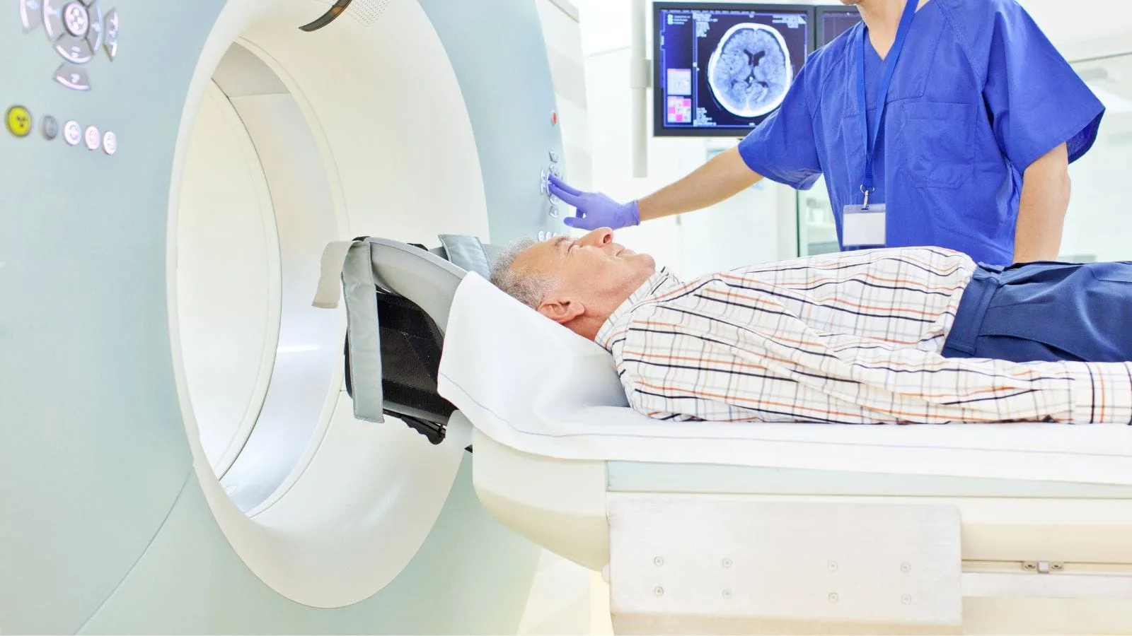
Diagnosis is usually made with brain MRI imaging. On MRI, hyperintense areas in the white matter can be observed, especially in T2 and FLAIR sequences. These areas suggest chronic ischemic changes.
The following elements are evaluated together when making a diagnosis:
- Detailed patient history
- Neurological examination
- Blood pressure measurement
- Blood tests (glucose, lipid profile, etc.)
- Advanced vascular imaging methods when necessary
The important point here is to determine whether the imaging finding is consistent with the patient’s clinical condition.
Does It Progress, Does It Cause Stroke?
The answer to this question varies from person to person. Small vessel disease is a process that can progress over time. In the presence of uncontrolled risk factors, new ischemic foci may develop. This can lead to small strokes called lacunar infarctions in some individuals.
However, in many patients where appropriate risk factor control is achieved, the process may remain stable. In other words, not every small vessel change necessarily results in severe neurological loss. The clinical course is influenced by variables such as blood pressure control, metabolic status, and lifestyle.
Treatment and Follow-Up Approach
There is no “single medication” for small vessel occlusion. The treatment approach is based on controlling factors that may increase underlying vascular damage.
General principles include:
- Regular monitoring and control of blood pressure
- Maintaining blood sugar balance
- Monitoring cholesterol levels
- Quitting smoking
- Regular physical activity
- Balanced nutrition
In some patients, antiplatelet (blood-thinning) therapy may be considered according to the physician’s evaluation; however, this decision is made based on individual risk analysis. The same approach does not apply to every patient.
When Should You Consult a Neurology Specialist?
If findings related to small vessel disease are indicated in the MRI report and the patient experiences any of the following complaints, a neurological evaluation is recommended:
- Progressive forgetfulness
- Difficulty in daily living activities
- Loss of balance
- Sudden onset neurological symptoms
In addition, if there are comorbid conditions such as hypertension or diabetes, regular follow-up is important.

Prof. Dr. Özgür Kılıçkesmez is one of Türkiye’s leading interventional radiology and neuroradiology specialists, focusing on minimally invasive endovascular treatments. His primary areas of expertise include aneurysm and stroke treatments, embolization procedures, non-surgical treatments of thyroid and parathyroid diseases, interventional oncology, and peripheral vascular interventions. He currently serves as the Head of the Interventional Radiology and Neuroradiology Department at Istanbul Memorial Göztepe Hospital.
Graduating from Istanbul University Cerrahpaşa Faculty of Medicine in 1997, Prof. Dr. Kılıçkesmez received advanced training in interventional radiology and oncology as a Clinical Fellow at London Guy’s & St Thomas’ and King’s College Hospital between 2012 and 2013. He earned the title of Professor in 2020 and, in the same year, became the Founding Interventional Radiologist at SBÜ Çam and Sakura City Hospital. He also holds the EBIR (European Board of Interventional Radiology) certification.
Prof. Dr. Kılıçkesmez has 94 international and 40 national publications and over 2500 citations. He serves as a reviewer for prestigious journals such as the American Journal of Roentgenology (AJR) and Diagnostic & Interventional Radiology. With his clinical and academic expertise, he is a nationally and internationally recognized authority in stroke treatment, brain aneurysm, embolization, tumor ablation, and minimally invasive treatments of thyroid/parathyroid diseases.







Vaka Örnekleri