


Disc extrusion is a spinal disorder where the inner nucleus of the intervertebral disc extends beyond the outer layer, compressing nearby nerves. It often causes severe back or leg pain and neurological symptoms.
Common causes of disc extrusion include degenerative disc disease, trauma, and excessive spinal strain. Risk factors such as aging, obesity, and repetitive heavy lifting increase the likelihood of occurrence.
Symptoms of disc extrusion may include sciatica, numbness, tingling, or weakness in the limbs. Severe cases can lead to mobility limitations or loss of bladder and bowel control, requiring urgent evaluation.
Treatment ranges from conservative management with physical therapy and medications to minimally invasive interventions or surgery. The approach depends on symptom severity and neurological findings.
| Medical Name | Disk Extrusion (Nucleus pulposus protruding beyond the outer layer) |
| Causes | Trauma, heavy lifting, sudden movements, age-related disc degeneration |
| Risk Factors | Advanced age, heavy physical activity, prolonged sitting, obesity, smoking |
| Common Symptoms | Low back/neck pain, radiating pain to the leg/arm (radiculopathy), numbness, tingling, muscle weakness |
| Diagnostic Methods | MRI imaging, physical examination, neurological tests |
| Observed Complications | Nerve root compression, sensory loss, risk of paralysis (in severe cases), loss of bladder-bowel control |
| Treatment Methods | Rest, physical therapy, painkillers, muscle relaxants, epidural steroid injections, nucleoplasty |
| Surgical Necessity | If there is severe pain, muscle weakness, or neurological loss: methods such as microdiscectomy, laminectomy |
| Prevention Methods | Proper lifting techniques, regular exercise, posture correction, weight control |
What Exactly Is Disk Extrusion?
Between the bones of our spine are discs that act like cushions. Their inner part is soft and jelly-like, while the outside is firmer and rubbery. Thanks to these discs, our spine can move and bear loads.
In disk extrusion, the soft inner part of the disc is forced outward through a tear in the outer layer. Imagine jam inside a pastry oozing out through a crack but still attached to the pastry. Disk extrusion is similar to this situation.
How Is Disk Extrusion Different From Other Herniations?
There are different degrees of herniation. The main feature that distinguishes disk extrusion from others is that the inner material of the disc passes completely through the outer layer but still remains connected to the disc.
In protrusion (bulging), which is a milder form, the outer layer is not torn but simply stretches outward.
In sequestration (fragmentation), which is a more advanced condition, the extruded material is completely detached and becomes a free fragment within the spinal canal.
Extrusion is generally more serious than bulging, as it is more likely to compress nerves. Knowing the type of herniation helps us plan the appropriate treatment.
How Does Disk Extrusion Occur?
Disk extrusion usually results from a combination of factors. Most commonly, the underlying process is degeneration—the gradual wear and tear of the disc over time. As we age, discs lose water, become less flexible, and more brittle, making the outer layer more susceptible to tears.
Additional mechanical stress can accelerate this process. Lifting heavy objects incorrectly, sudden or awkward movements, repetitive bending or twisting, or trauma such as a fall can cause a tear in the weakened outer layer. Once a tear occurs, the soft inner part pushes outward.
What Is the Role of Inflammation in Disk Extrusion?
The extruded disc material not only compresses nerves mechanically, but is also recognized as a foreign substance by the body, which triggers inflammation. This inflammation releases chemicals that irritate nerves and increase pain.
However, inflammation also has a positive side. By attracting the body’s defense cells to the area, it helps clean up the herniated material. So inflammation both causes pain and is part of the healing process. We take this balance into account during treatment.
What Are the Main Causes of Disk Extrusion?
There are usually two main causes underlying disk extrusion. The first is the natural aging and weakening of discs, which is the most common situation.
The second is sudden or repeated loading of the spine. For example, traffic accidents, falls from heights, or repetitive heavy lifting and working in poor positions can cause a disc tear. Often, these two factors act together; a previously worn disc may tear with even a minor strain.
What Are the Risk Factors for Disk Extrusion?
- Genetic predisposition: family history of herniation increases risk.
- Excess weight: especially increases the load on lumbar discs.
- Occupation: those who lift heavy, bend and twist frequently, or sit/drive for long periods are at higher risk.
- Smoking: impairs nutrition of the discs and accelerates degeneration.
- Sedentary lifestyle: weakens the muscles supporting the spine.
- Poor posture habits: place uneven loads on the discs.
- Certain diseases: conditions like diabetes may also increase risk.
Some risk factors are modifiable (weight, smoking, exercise), while others are not (age, genetics). You can reduce your risk by focusing on modifiable factors.
Does Disk Extrusion Always Cause Symptoms?
No, not every disk extrusion causes symptoms. Sometimes, a disk extrusion is detected incidentally on spinal imaging (such as MRI) performed for another reason, and the person has no complaints. This is called an “asymptomatic herniation.”
When symptoms do occur, it is usually because the extruded disc fragment is compressing or chemically irritating the nerve roots. The symptoms depend on the location of the herniation (lumbar or cervical) and which nerve is affected.
What Symptoms Does Lumbar Disk Extrusion Cause?
Lumbar herniations (lumbar disk extrusion) most commonly occur in the lower back. The most typical symptom is sciatica, which usually starts from the hip and radiates down the back of the leg, sometimes to the foot. The pain may feel burning, stabbing, sharp, or electric-like and is usually unilateral. Coughing or sneezing can worsen the pain.
Other symptoms include low back pain, numbness or tingling in the leg or foot, weakness in affected muscles (such as foot drop), and reduced reflexes.
What Are the Symptoms of Cervical Disk Extrusion?
Cervical herniations (cervical disk extrusion) mostly occur between the middle and lower cervical vertebrae. Neck pain is common and may increase with head movement.
The most prominent complaint is usually pain radiating to the arm. Pain can start from the shoulder and extend to the arm, hand, and fingers. Which finger is affected can help determine which nerve is involved. Numbness, tingling, and muscle weakness (such as difficulty holding objects) in the arm, hand, or fingers are common. Sometimes pain is also felt between the shoulder blades.
What Are the Emergency Symptoms in Disk Extrusion?
Certain symptoms require immediate medical attention—these are called “red flag” symptoms.
One example is Cauda Equina Syndrome, which may occur with lumbar herniations. Symptoms include progressive leg weakness, loss of bladder or bowel control, and numbness in the genital or anal region. This requires emergency surgery to prevent permanent nerve damage.
Sudden or rapidly worsening muscle weakness in the arm or leg also requires urgent evaluation. If you experience these symptoms, seek medical care immediately.
What Examinations Are Performed to Diagnose Disk Extrusion?
Diagnosis starts with a detailed history: when and how symptoms began, their exact location, severity, and what worsens or relieves them. Your medical history and overall condition are also reviewed.
A physical exam follows. Posture and gait are observed, the spine is palpated for tenderness or muscle spasm, and the range of neck and lower back movement is evaluated.
The most important is the neurological exam, which checks muscle strength, sensation (touch, pain), and reflexes in the arms and legs. Specific tests, such as the straight leg raise for lumbar herniation or maneuvers for cervical herniation, are used to assess nerve involvement.
What Imaging Methods Are Used to Diagnose Disk Extrusion?
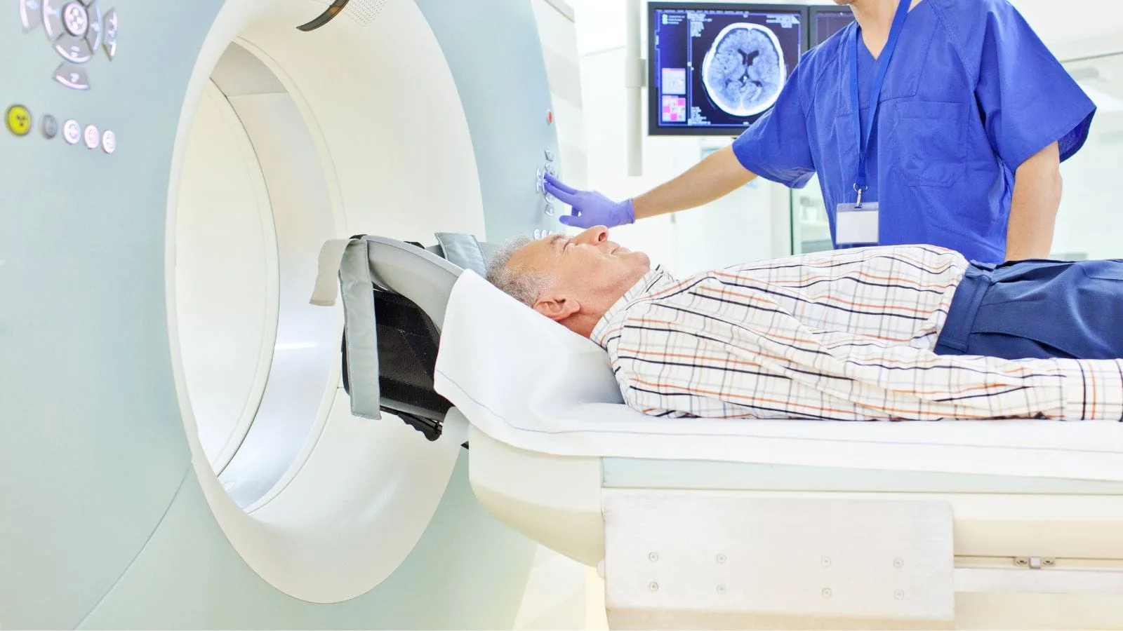
- Magnetic Resonance Imaging (MRI) is the gold standard, showing discs, nerves, and the spinal cord clearly. It reveals the location, size, type of herniation, and the extent of nerve compression, providing the most valuable information for planning treatment.
- Computed Tomography (CT) is more detailed for bone structures and is used if MRI is not possible or if additional bone problems are suspected.
- X-ray does not show the herniation but can be used initially to rule out bone problems such as fractures or degeneration.
Remember, finding a herniation on imaging does not always mean it is the cause of your complaints. Diagnosis combines your symptoms, examination findings, and imaging results.
How Are Injection Therapies Done for Disk Extrusion?
If pain is severe, epidural steroid injections may be used. In this procedure, a cortisone medication is injected into the epidural space around the nerve at the site of herniation, usually under X-ray or ultrasound guidance.
The aim is to reduce strong inflammation and swelling around the nerve to relieve pain. These injections do not remove the herniation but help control pain.
When Is Surgery Needed for Disk Extrusion?
Surgery is generally a last resort and considered in certain cases. If a person cannot get relief from severe pain despite adequate conservative treatment (usually at least 6 weeks to 3 months), surgery may be an option.
If there is progressive and worsening muscle weakness in the arm or leg, or emergency symptoms such as Cauda Equina Syndrome (loss of bladder/bowel control), surgery becomes unavoidable to prevent permanent nerve damage. The decision for surgery is made by considering your complaints, examination findings, and imaging together.
Are There Risks to Disk Extrusion Surgery?
As with all surgical procedures, disk extrusion surgery carries potential risks. These include infection, bleeding, damage to the nerve root or spinal cord covering (usually a low risk), leakage of spinal fluid, and general anesthesia risks.
There is also the possibility of insufficient pain relief or recurrence of the herniation at the same or another level, even after surgery. These risks are generally low, and surgeons take all necessary precautions to minimize them. However, it is important to discuss these potential risks with your doctor before surgery.
Can the Herniated Disk Heal on Its Own in Disk Extrusion?
Yes, interestingly, this is possible and quite common. The spontaneous regression of herniated disc material (shrinking or disappearing on its own) can occur thanks to the body’s natural defense and clearing mechanisms.
Especially in cases where the fragment is completely separated (sequestration) or extruded but still attached, the likelihood of regression is higher compared to milder bulging herniations. Larger herniations are even more likely to shrink. This process usually takes months, but shows that the body can heal herniations on its own. During this time, injection therapies may be more appropriate if pain worsens, but the choice depends on the individual and should be discussed with your doctor.
What Factors Affect Recovery in Disk Extrusion?
How well and how quickly a person recovers from disk extrusion depends on many factors. The type and size of the herniation (as mentioned, larger extrusions are more likely to shrink), duration of symptoms, and initial severity of muscle weakness are important.
Other factors include age, general health (such as the presence of diabetes), psychological state (resilience, optimism), whether the person smokes, and most importantly, adherence to treatment (regular exercise, following advice). Recovery depends not only on the herniation, but the person as a whole.
Why Is Posture Important in Preventing Disk Extrusion?
How we stand, sit, or sleep directly affects the load on our spine. Poor posture habits place constant and uneven loads on the discs, promoting degeneration and herniation.
Supporting the back when sitting, distributing weight evenly when standing, choosing a sleeping position and mattress that maintain the natural curvature of the spine, and organizing the work environment ergonomically (chair, desk, screen height) help protect the spine during long hours. Good posture is a simple yet effective way to reduce the risk of herniation.
What Is the Role of Exercise in Preventing Disk Extrusion?
Regular exercise plays a key role in preventing disk extrusion. Exercises that strengthen the deep core and back muscles supporting the spine reduce the load on the spine and increase its stability. Stabilization exercises such as planks and bridges are very effective.
Flexibility exercises (stretching) help maintain the mobility of the spine and related muscles. Aerobic activities that do not overload the spine, such as walking, swimming, or cycling, both improve general health and help nourish the discs. A strong and flexible body is one of the best defenses against herniation.
What Lifestyle Changes Can Prevent Disk Extrusion?
In addition to posture and exercise, general lifestyle habits affect disc health. Maintaining an ideal weight is crucial, as excess weight increases pressure on the lumbar discs.
Quitting smoking is also critical. Smoking reduces blood flow to the discs, accelerating degeneration and making healing more difficult.
Drinking enough water and having a generally healthy and balanced diet are important for tissue health. Learning proper lifting techniques and avoiding sudden, awkward movements in daily life are simple steps to reduce risk. These changes not only prevent herniation, but also improve your overall health.

Prof. Dr. Özgür Kılıçkesmez is one of Türkiye’s leading interventional radiology and neuroradiology specialists, focusing on minimally invasive endovascular treatments. His primary areas of expertise include aneurysm and stroke treatments, embolization procedures, non-surgical treatments of thyroid and parathyroid diseases, interventional oncology, and peripheral vascular interventions. He currently serves as the Head of the Interventional Radiology and Neuroradiology Department at Istanbul Memorial Göztepe Hospital.
Graduating from Istanbul University Cerrahpaşa Faculty of Medicine in 1997, Prof. Dr. Kılıçkesmez received advanced training in interventional radiology and oncology as a Clinical Fellow at London Guy’s & St Thomas’ and King’s College Hospital between 2012 and 2013. He earned the title of Professor in 2020 and, in the same year, became the Founding Interventional Radiologist at SBÜ Çam and Sakura City Hospital. He also holds the EBIR (European Board of Interventional Radiology) certification.
Prof. Dr. Kılıçkesmez has 94 international and 40 national publications and over 2500 citations. He serves as a reviewer for prestigious journals such as the American Journal of Roentgenology (AJR) and Diagnostic & Interventional Radiology. With his clinical and academic expertise, he is a nationally and internationally recognized authority in stroke treatment, brain aneurysm, embolization, tumor ablation, and minimally invasive treatments of thyroid/parathyroid diseases.






Vaka Örnekleri