


Bleomycin electrosclerotherapy (BEST) in interventional radiology is an advanced medical method that treats complex vascular and lymphatic system abnormalities completely without surgery. This technique combines the power of a special medication applied to the tissue with instantaneous electrical pulses to provide permanent healing at the cellular level.
Vascular malformations in anatomical regions where surgical procedures are considered risky can be safely eliminated using this method without disrupting the integrity of the patient’s tissues. Large incisions or permanent scars are absolutely unnecessary.
Performed under the guidance of advanced medical imaging devices, these procedures focus directly on the affected area with pinpoint precision. Only the diseased structure is targeted without damaging healthy organs or surrounding tissues.
Offering patients a very short recovery period, this innovative approach is one of today’s most effective non-surgical solutions for vascular problems that significantly reduce quality of life.
What Is the Bleomycin Electrosclerotherapy (BEST) Method?
The foundation of this treatment method lies in the harmonious interaction between biophysics and pharmacology. To better understand this process, it is useful to first take a closer look at the building blocks of our bodies, namely our cells. Every cell in our body is surrounded by a very tight, lipid-based, double-layered membrane that protects it from external influences and determines which substances can enter and leave it. Under normal circumstances, this membrane acts as an extraordinary security barrier that ensures the cell’s survival. Although the special medication called bleomycin used in the treatment is highly effective at stopping the proliferation of diseased cells that form the vascular abnormality, its large molecular structure makes it difficult to penetrate this tight cell membrane on its own. If the medication merely comes into contact with the outer surface of the cell, the expected therapeutic effect is unlikely to occur.
This is precisely where reversible electrical permeability technology, which lies at the heart of the treatment, comes into play. During the procedure, high-voltage electrical pulses lasting only a millionth of a second are delivered to the cell membrane through thin needles placed inside the abnormal vascular mass. This electrical stimulation helps create temporary pores or openings on the nanometer scale, meaning they are too small to be seen with the naked eye, in the protective barrier surrounding the cell. This process is medically known as electroporation. Through these microscopic openings in the cell membrane, the medication waiting outside begins to enter the cell within seconds at a much higher concentration than would normally be possible.
As soon as the electrical current stops, the cell membrane generally closes these tiny openings on its own within seconds without sustaining permanent damage and returns to its original intact structure. This is why the process is called reversible; the cell is not physically destroyed and continues to maintain its structural integrity. However, the medication that has now reached the nucleus, the very center of the diseased cell, is trapped inside. The medication begins to create breaks in the structural components of the diseased vascular cell, preventing it from dividing and multiplying. It gives the cell a programmed and completely natural command to cease its activity. Over time, the body’s own immune system clears away these diseased cells that have reached the end of their lifespan, allowing the swollen vascular mass in that area to permanently contract and regress.
What Does Vascular Locking Mean During Bleomycin Electrosclerotherapy (BEST), and Why Is It Important?
Blood continuously flows through the vessels at a certain speed, which is a natural process. When any medication is injected into a blood vessel during conventional needle treatment, the flowing blood can quickly wash the medication away from the area. Because the medication cannot remain in the affected region long enough, its effectiveness may decrease, and it may reach unintended areas as it spreads throughout the body through the bloodstream. One of the greatest advantages offered by this method is the vascular locking effect created during the procedure.
The very brief electrical pulses applied not only create openings in cell membranes but also cause the muscle fibers in the vessel walls to contract strongly and reflexively. This sudden contraction substantially stops blood flow inside the abnormal vascular mass at that moment. The vessel virtually closes in on itself, creating a locking mechanism and effectively isolating itself.
The role of this locking effect in treatment is highly valuable. The medication administered into the affected area has little opportunity to escape into the systemic circulation, meaning other parts of the body, the heart, and the lungs. The medication remains largely trapped within the target tissue where it is intended to act. Since the washing effect of blood is eliminated, the medication has sufficient time and favorable conditions to penetrate the cells lining the inner surface of the diseased vessels.
The main advantages provided by vascular locking are listed below.
- Trapping the medication within the targeted tissue
- Protecting the body’s other organs
- Significantly increasing medication effectiveness
- Reducing the required medication dose
For Which Diseases and Conditions Is Bleomycin Electrosclerotherapy (BEST) Preferred?
In clinical practice, this treatment method is primarily considered for structurally complex groups of lesions, particularly those known as slow-flow vascular abnormalities. These abnormalities are mainly divided into two groups, venous disorders and lymphatic system disorders, with different structural problems observed in each group.
Venous abnormalities consist of dilated, spongy, and malformed vascular masses. Because blood flow within these diseased vessels slows considerably, localized blood clotting may occur. This condition can cause episodes of pain that profoundly affect the patient’s daily quality of life. By entering these dilated vascular pools, the method initiates a strong contraction process within them and supports closure of the abnormal vascular pathway. It is a particularly helpful option for widespread problems that have infiltrated muscle tissue.
It is also safely used in arteriovenous malformations (AVMs), which are predominantly arterial vascular anomalies, particularly in areas such as the tongue, lips, jaw, ears, and fingers where embolization is impossible or very risky, as well as in cases where the malformation affects very large areas and in recurrent, persistent AVMs.
Lymphatic system abnormalities arise from developmental defects in the body’s fluid-balancing system and appear as clusters of fluid-filled cysts. If the cysts are large, they may respond well to conventional needle treatments. However, the real challenge involves microscopic lymphatic abnormalities in which tens of thousands of tiny cysts spread between tissues like a sponge. It is physically almost impossible to insert a needle into each of these tiny cysts individually, and conventional medications have great difficulty penetrating them. Because electrical current provides extensive and homogeneous cellular permeability throughout the tissue spaces, the medication can reach even the walls of these microscopic cysts and selectively target the entire spongy structure.
The main complaints caused by these diseases that lead patients to seek treatment are as follows.
- Clearly visible swellings
- Purple or bluish discoloration of the skin
- Localized and recurrent pain
- Episodes of clotting within blood vessels
- Deformities and postural abnormalities
Why Is Bleomycin Electrosclerotherapy (BEST) Used Instead of Traditional Treatments?
In extremely complex anatomical regions such as the head and neck, or in areas where movement function is critical, such as the hand, surgically cutting out and removing abnormal vascular masses is not always an easy process. These problematic tissues often surround normal muscles, vital nerves, and healthy arteries, infiltrating the tissues almost like ivy. During a surgical procedure, damage to a nerve responsible for the patient’s facial expressions or removal of a lesion on the tongue may put speech and swallowing functions at risk. Moreover, even after surgical intervention, the possibility of the disease becoming noticeable again over the years due to diseased cells remaining at the microscopic level often persists.
On the other hand, traditional needle treatments that use only chemical substances also present certain challenges. These chemicals work by producing a caustic and destructive effect on the vessel wall. However, this chemical effect cannot always be contained. The medication may sometimes leak outside the vessel and affect adjacent healthy muscle tissue or nerves, causing unwanted damage to surrounding tissues.
The distinguishing feature of this advanced method compared with traditional treatments is that it does not generate heat in the tissues. The electrical current does not burn, freeze, or dissolve the tissue like a chemical acid. It only creates temporary openings in cell membranes. Therefore, nerves, the structural scaffolds forming the surrounding connective tissue, and healthy neighboring arteries do not undergo physical destruction and can often be safely preserved. The medication focuses only on the targeted diseased cells lining the inner surface of blood vessels that divide rapidly. Because tissue architecture is preserved, the aim is to maintain the patient’s existing functions and achieve a recovery process that generally leaves fewer visible marks. It stands out as an important option even for individuals who have previously undergone surgery or have not benefited sufficiently from standard treatments.
How Do the Preparation Stages Progress Before Bleomycin Electrosclerotherapy (BEST)?
The success of treatment depends greatly on the meticulous mapping and preparation performed before the procedure, much like planning a detailed journey. At this stage, advanced imaging methods are used to examine the inside of the body in detail without surgery and determine the location and structure of the problem. This is like giving the specialist performing the procedure a flashlight to find their way in the dark.
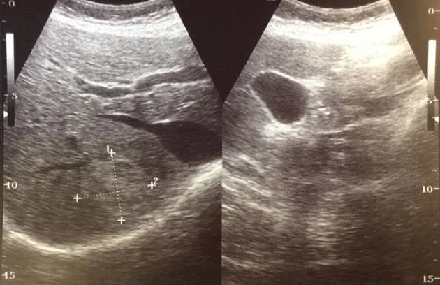
Before the procedure, ultrasound is used to carefully evaluate the boundaries of the abnormal vascular mass, the extent of its spread, the speed of blood flow within it, and its close relationship with surrounding healthy tissues. Ultrasound is a device that displays the movement of blood within vessels in real time and is very useful for understanding the dynamics of the area. After superficial mapping is completed, an MRI is generally performed to determine how far the problem extends into deeper tissues, around muscles, or around bones. MRI provides a detailed map of the affected area. This mapping makes it possible to plan where and at what angle the needles will be positioned during the procedure.
The main preparation steps planned before the treatment day include:
- Mapping with ultrasonography
- Review of MRI imaging
- Blood tests
- Evaluation of respiratory functions
- Examination by an anesthesiologist
How Is Bleomycin Electrosclerotherapy (BEST) Performed Step by Step in a Hospital Setting?
To ensure patient comfort and create a safe environment, this procedure is generally performed in a fully equipped angiography suite or operating room through the cooperation of the anesthesia team and relevant specialists. One of the subjects patients are curious about is the sensation electricity may create in the body. Since electrical stimulation can naturally cause involuntary muscle contractions, care is taken not to perform the procedure while the patient is awake. General anesthesia is administered, and special medications are given to relax the muscles. Thus, the patient remains asleep throughout the procedure and generally feels nothing; the operation is performed in a motionless and controlled environment.
One of the most important safety steps of the procedure is synchronization with the heart’s rhythm. The human heart operates through its own electrical system, and care is taken to ensure that a strong externally delivered electrical stimulus does not affect this system. To achieve this, the device is connected directly to the patient’s heart monitor. The device monitors every heartbeat and identifies the very brief period when the heart is electrically at rest, delivering its own pulse at precisely that moment. Especially in areas around the chest or neck, this synchronization provides a valuable safety measure.
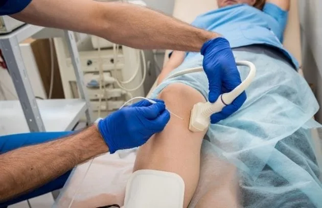
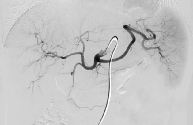
After the patient is safely anesthetized, the specialist uses an ultrasound device as a guide to place special thin needles directly into the affected area. If there is a fluid-filled structure belonging to the lymphatic system, the fluid inside is carefully drained first. Then, a contrast fluid is administered, and the connections between the abnormal vessels and the body’s blood circulation pathways are monitored in real time. After the blood outflow pathways are temporarily restricted, the prepared medication solution is injected into the lesion. Following a short waiting period to allow the medication to spread between the tissues, very brief electrical pulses begin to be delivered through the inserted needles. The current is applied systematically, ensuring that the medication remains within the intended area.
How Are Medication Dosage and Safety Precautions Planned in Bleomycin Electrosclerotherapy (BEST)?
Since every patient’s anatomy, the size of the problem, and bodily dynamics differ, the details of the treatment are also personalized. The amount of medication to be used is calculated very carefully according to the patient’s body weight, the extent of the affected area, and its structure within the tissue. While much lower doses are sufficient for small affected areas, the amount and method of medication administration are adjusted according to the need in conditions involving larger areas.
When people hear the names of certain medications, they may think of the high-dose and exhausting processes used in cancer treatment. It is known that the medication called bleomycin may cause long-term strain on the lungs when it spreads throughout the body at high doses. However, the doses used when applying this interventional radiology method are considerably below the limits used in systemic treatments. Only a very small fraction of the safe dose limit that can normally be administered to a person is considered in a single session. Moreover, thanks to the vascular locking technique applied, the medication is prevented from spreading throughout the body and is kept within the targeted area as much as possible. Therefore, the risk of placing strain on the lungs or other organs remains very low.
Nevertheless, taking precautions is essential in healthcare. To minimize even the smallest risks, anesthesiologists conduct careful monitoring during the procedure. Since high concentrations of inhaled oxygen while the medication is in the body may sometimes increase lung sensitivity, the oxygen concentration delivered to the patient through the anesthesia machine is deliberately kept below a certain level. This small but highly scientific precaution contributes significantly to a smoother treatment process.
The main safety precautions taken during the procedure are listed below.
- Reducing the oxygen concentration below thirty percent
- Synchronizing with the heart using an ECG device
- Regular blood pressure monitoring
- Close monitoring of the respiratory tract
What Can Patients Expect During Recovery After Bleomycin Electrosclerotherapy (BEST)?
When the patient is awakened after the procedure, they may be discharged from the hospital the following day and return home if their general health condition permits. However, certain expected reactions may begin in the treated area of the body as a natural consequence of the medication and the method applied. During the first days after the procedure, noticeable swelling, tenderness, and pain associated with changes inside the blood vessels in the treated area are quite normal.
This is generally not a sign that something has gone wrong; on the contrary, it indicates that the applied method and the body’s immune system have begun responding to the diseased cells. This temporary discomfort and swelling can often be easily controlled with painkillers recommended by the physician and localized cold applications. The complaints generally decrease gradually over several weeks, giving way to permanent tissue healing.
In addition, another temporary condition may occur at the skin level. In vascular problems located very close to the skin, the electrical current used may produce a mild effect at the needle insertion points. Furthermore, a very small amount of medication may be present immediately beneath the skin. As a result, temporary darkening of the skin resembling fine scratches or marks may occur. Specialists calculate the electrical fields very carefully and take protective measures for the skin to minimize this visible effect. The vast majority of such skin marks tend to fade and disappear over time.
The usual conditions patients may experience during the recovery period are generally as follows.
- Edema in the treated area
- Mild or moderate tenderness
- Temporary linear discoloration of the skin
- Localized sensitivity to touch
What Are the General Results and Success Rates of Bleomycin Electrosclerotherapy (BEST)?
When observations and clinical follow-ups are examined, this method appears capable of producing encouraging results, particularly in resistant cases. In conventional surgery or needle treatments, it may not always be easy to reach every part of the tissue equally or eliminate the problem without leaving anything behind. For this reason, diseases may occasionally recur.
However, the findings obtained with this method, which combines electrical permeability with a special medication, are quite promising for patients. In many individuals who have previously tried different treatments or whose conditions were considered difficult to treat surgically, imaging performed months after the procedure shows that the volume of diseased tissue has decreased significantly and that the lesion has largely regressed.
Beyond volumetric reduction, very good results are obtained in managing complaints such as pain, clotting, or a feeling of heaviness that directly affect patients’ quality of life. In a large proportion of patients, these uncomfortable symptoms decrease noticeably. Particularly in problems located close to the respiratory tract or areas involved in speech or swallowing, the aim is to preserve healthy tissue while reducing only the problematic portion, allowing patients to continue their daily lives, often without losing their existing functions.
What Is the Role of the Interventional Radiologist in Bleomycin Electrosclerotherapy (BEST)?
These types of vascular abnormalities, hidden deep within our bodies and located between muscles or around critical nerves, are conditions that require specialized expertise to overcome. Understanding these structures simply by looking from the outside or eliminating them with conventional methods without affecting surrounding tissues is often quite challenging. This is where interventional radiology, a modern branch of medicine, comes into play.
An interventional radiologist is a specialist who can visualize the inside of the body using advanced medical imaging devices without the need for surgical incisions and simultaneously use these images to deliver treatment directly to the center of the problem through very thin needles. Successful application of this treatment depends on correctly positioning the needles, safely restricting blood flow pathways, and calculating the energy delivered to the body with great precision. This entire process falls within the expertise of interventional radiology, which combines the ability to use advanced technology with the skill to perform delicate manual procedures.
In cases involving slow-flow vascular abnormalities, especially when previous treatments have not provided sufficient benefit or surgical intervention has been considered difficult, this method emerges as an innovative and effective treatment option for patients. When this approach, which aims to achieve healing at the cellular level while focusing on preserving healthy tissues, is evaluated with careful and meticulous planning under the guidance of a specialist who will manage the process, it can become one of the most valuable steps toward achieving lasting relief.








Vaka Örnekleri