


Advanced imaging techniques in the diagnosis of erectile dysfunction provide precise evaluation of vascular, neurogenic, and structural factors, enabling targeted and evidence-based management. Modalities such as Doppler ultrasound and MRI offer objective data on penile hemodynamics and tissue integrity.
Penile Doppler ultrasound assessment of erectile dysfunction focuses on arterial inflow and venous occlusive function, allowing detailed measurement of peak systolic velocity and end-diastolic flow. This technique is widely used due to its accessibility, real-time capability, and high diagnostic value in vascular etiologies.
Magnetic resonance imaging in erectile dysfunction diagnosis provides high-resolution visualization of soft tissues, including the corpora cavernosa and surrounding neurovascular structures. Functional MRI techniques further enhance the understanding of central and peripheral mechanisms contributing to erectile impairment.
Computed tomography angiography for erectile dysfunction enables comprehensive mapping of pelvic and penile vasculature, particularly in complex or post-traumatic cases. This advanced modality supports surgical planning and improves diagnostic accuracy when conventional imaging methods yield inconclusive findings.
What Is Erectile Dysfunction (Erectile Dysfunction) and What Are Its Vascular Causes?
A healthy erection is actually proof of how perfectly engineered the human body is. We can compare this process to inflating a car tire. For the tire to be firm and ready for use, two basic things are needed: a sufficient amount of air pumped inside and a solid valve that prevents the air from escaping.
In our body, this process works as follows: When sexual stimulation occurs, signals from the brain cause the arteries carrying blood to the penis to dilate. This is like the compressor starting to pump air into the tire. Blood rapidly fills the spongy tissues inside the penis called the “corpus cavernosum.” As these tissues fill with blood and expand, they press against the tough, non-elastic sheath surrounding the penis called the “tunica albuginea.” This pressure compresses and closes the veins responsible for draining blood back from the penis. This is the moment when the valve of the tire closes. The blood that enters cannot escape, pressure increases, and an erection occurs.
In the condition known as “venous leak,” the problem lies in this valve mechanism. Although enough blood flows into the penis through the arteries, the veins cannot compress and close properly. Because the valve of the tire is faulty, the air that enters immediately leaks out. As a result, the penis either cannot achieve sufficient rigidity at all, or the achieved erection is lost very quickly. This is one of the most common physical causes of erectile dysfunction.
So why does this “valve mechanism” fail? Some of the main vascular causes underlying venous leak are as follows:
- Weaknesses in the structure of the tunica albuginea (sheath)
- Reduced ability of the muscles within the spongy tissue to relax
- Congenital abnormal vascular connections
- Previous trauma or accidents
- Tissue changes related to Peyronie’s disease
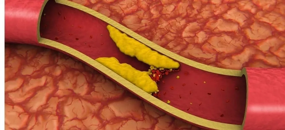
- Damage caused by long-standing diabetes or atherosclerosis
Correctly identifying these causes is vital for the success of treatment, and this is exactly where advanced imaging comes into play.
When Are Advanced Imaging Methods Needed in Erectile Dysfunction?
When you consult a physician with complaints of erectile dysfunction, the process usually proceeds step by step. First, a detailed medical history is taken, and questions are asked about your lifestyle, habits, and sexual life. This is followed by a physical examination and sometimes blood tests to check hormone levels.
In most cases, the first treatment option is oral medications that increase blood flow to the penis. These medications provide an effective solution for millions of men. However, some patients may not benefit sufficiently from this treatment. In such cases, it becomes necessary to delve deeper to find the answer to the question, “Why isn’t it working?”
When drug therapy fails, the likelihood of an underlying vascular problem increases. The first and most important test performed to clarify this suspicion is Penile Color Doppler Ultrasonography. During this test, a medication that induces an artificial erection is injected into the penis, and blood flow within the penis is monitored in real time using ultrasound. This test provides us with two critical pieces of information.
- Whether sufficient blood is reaching the penis through the arteries at an adequate speed
- How quickly blood is leaking back through the veins
If Doppler ultrasonography shows that arterial flow is normal but there is significant venous leakage, then you become a candidate for advanced imaging methods. These methods are used not only to confirm the presence of leakage but also to reveal where it originates from, which veins are responsible, and the pathway it follows. For the Interventional Radiologist planning the treatment, this means having a detailed map in hand before taking the next step.
What Is Conventional Cavernosography and Why Is It No Longer Sufficient?
For many years, cavernosography was considered the only and most reliable method—the “gold standard”—for diagnosing venous leak. Its basic principle is to inject a contrast agent visible on X-ray into the spongy tissues of the penis and observe under live X-ray imaging (fluoroscopy) how and how quickly this dye passes into the veins.
This method was quite successful in demonstrating whether a leak was present. If the dye appeared in the veins outside the penis during erection, this was definitive proof of leakage. However, with the advancement of technology and treatment methods, a major shortcoming of conventional cavernosography became evident: it is two-dimensional (2D).
Imagine trying to understand a large city with a complex highway network, bridges, overpasses, and underpasses. Conventional cavernosography is like looking at a black-and-white aerial photograph of this city taken from a single angle, where all roads and buildings overlap in a blurry, single plane. You may see movement or a “leak” in the photo, but it is impossible to say exactly which road it starts from, which bridge it passes under, and where it ends. The complex vascular structure of the penis and pelvic region is very much like this city. In conventional cavernosography, the vein responsible for the leak may be hidden behind the pelvic bones, or multiple veins may overlap and appear as a single vessel.
This was also the main reason why venous leak surgeries performed in the past often failed. Surgeons would ligate only the most prominent vein visible on this inadequate map, while missing other hidden veins that were actually responsible for the leak. This caused the problem to recur after a short time.
The main reasons why conventional cavernosography is insufficient for treatment planning today are as follows:
- Two-dimensional imaging principle
- Superimposition of structures and vessels
- Inability to clearly identify the source of the leak
- Failure to provide a roadmap for treatment
- Higher radiation dose compared to modern methods
- Longer and more cumbersome procedure for the patient
For these reasons, conventional cavernosography has now been replaced by modern technologies that show every street of the city in three dimensions.
CT Cavernosography: How Does It Create a Three-Dimensional Map of Vascular Leakage?
CT cavernosography (Computed Tomography Cavernosography) is the modern and most effective diagnostic tool developed to eliminate all the shortcomings of the conventional method. While the traditional technique offers a single blurry aerial photograph, CT cavernosography provides an interactive three-dimensional (3D) digital map that shows every centimeter of the highway network, every bridge, every tunnel, and every street, allowing you to rotate and view it from different angles.
This technology uses a computed tomography scanner that creates cross-sectional images of the body. During the procedure, the penis and pelvic region are scanned in hundreds of slices with millimetric precision. These slices are then combined by powerful computer software to create a flawless 3D model of the vascular network. Thanks to this model, the problem of image superimposition is completely eliminated. Each vessel can be clearly followed independently from bones and other tissues, from its origin to its endpoint.
CT cavernosography is performed by an experienced Interventional Radiologist in a modern CT suite, and its steps are quite comfortable for the patient.
- Preparation: The procedure area is cleaned, and a small local anesthetic injection is administered at the needle entry point to numb the area. This ensures that no pain is felt during the procedure.
- Induction of Erection: As with other methods, a medication that induces an erection is injected into the penis with a fine needle to best observe the venous leak mechanism. The procedure continues once sufficient rigidity is achieved.
- Administration of Contrast Material: Through a second fine needle placed into the other half of the penis, a harmless iodine-based contrast agent is administered at a controlled rate using an automatic injector. This allows all vessels to appear bright and clearly visible during the CT scan.
- CT Scan: While the contrast material is being administered, the patient lies on the CT table, and the scanner completes the entire scan within a few seconds. This part of the procedure is very fast.
- Three-Dimensional Modeling: After the scan, the real magic begins. The hundreds of cross-sectional images obtained are processed at specialized workstations. The Interventional Radiologist creates angiographic maps of the vessels and 3D models that can be rotated from any desired angle.
Thanks to this detailed map, all leakage points are identified with near 100% accuracy before treatment. This is the most important step in guaranteeing success, especially when planning minimally invasive treatments such as endovascular embolization.
The main advantages of CT cavernosography are as follows:
- Excellent three-dimensional imaging
- Complete elimination of the superimposition problem
- Very high accuracy in identifying leakage points
- Providing a detailed roadmap for treatment
- Extremely fast acquisition time
- Availability in many centers today
In Which Special Situations Is MR Cavernosography Used and What Additional Information Does It Provide?
MR cavernosography (Magnetic Resonance Cavernosography) is another powerful and specialized imaging method used in the diagnosis of venous leak. While CT cavernosography is excellent at mapping vascular anatomy, MR cavernosography goes one step further by providing different and highly valuable information.
MR cavernosography has two well-known and important advantages: it does not involve radiation and it can display soft tissues with exceptional detail. The absence of radiation makes it a very attractive option, especially for younger patients with erectile dysfunction who wish to minimize lifetime radiation exposure.
However, the real distinguishing feature of MRI is its soft tissue contrast. If we consider CT cavernosography as a technology that maps the vascular road network, MR cavernosography both maps this network and evaluates the quality of the ground on which the roads are built (that is, the penile tissue itself). Erection depends not only on healthy vessels but also on the spongy tissue that fills with blood being healthy and elastic. MRI can evaluate the health of this tissue using special imaging sequences called T2-weighted sequences.
MR cavernosography is invaluable in evaluating conditions such as the following:
- Corporal Fibrosis: A condition in which elastic spongy tissue inside the penis is replaced by stiff, nonfunctional connective tissue due to causes such as diabetes, trauma, or previous priapism (prolonged painful erection). This prevents the tissue from filling with blood.
- Peyronie’s Disease: A condition characterized by the formation of hard plaques within the penis, causing pain and curvature. MRI clearly shows the location and size of these plaques.
- Post-Traumatic Damage: Evaluates the extent of damage to internal penile tissues following an accident or injury.
- Integrity of the Tunica Albuginea: Examines the thickness and integrity of the sheath surrounding the penis, which plays a key role in the venous leak mechanism.
This capability of MRI allows us to answer a critical question in treatment planning: “Even if we successfully close the venous leak, is the underlying tissue functional enough to produce a healthy erection?” If widespread fibrosis is detected on MRI, it can be anticipated that embolization alone may not be sufficient, and more permanent solutions such as a penile prosthesis may be recommended. This protects the patient from a potentially unsuccessful procedure.
However, MR cavernosography also has some disadvantages. The imaging time is considerably longer than CT, and the patient must remain still throughout the procedure. It is also more costly and available in fewer centers.
Which Method Is Most Suitable for Me: Cavernosography, CT, or MRI?
Choosing among these three methods is like a tailor making a custom suit specifically for you. Each patient’s situation differs in terms of age, overall health profile, and treatment goals. Therefore, the choice is made on an individual basis by an Interventional Radiologist. However, to outline a general framework:
Conventional Cavernosography: It now has only historical significance and should not be used for modern treatment planning. Its insufficient diagnostic information puts treatment success at risk.
CT Cavernosography: It is the gold standard for the vast majority of patients for whom endovascular embolization treatment is planned. It offers the ideal balance of speed, accuracy, accessibility, and cost-effectiveness. It provides the clearest possible roadmap required before treatment.
MR Cavernosography: It is used in more special and selected cases.
- Young patients who wish to avoid radiation exposure.
- Patients with severe allergy to iodine-based contrast agents or with renal failure.
- Complex cases in which an underlying tissue problem (fibrosis, Peyronie’s disease) is suspected and tissue health also needs to be evaluated.
The final decision is made by considering all these factors in order to determine the most appropriate and successful treatment pathway for you.
How Do Imaging Results Shape Treatment and Increase Success?
The detailed anatomical maps obtained from advanced imaging methods are not merely diagnostic reports; they are action plans that form the foundation of venous embolization (vessel occlusion) procedures. This minimally invasive procedure is performed by Interventional Radiologists, usually by accessing through the groin or directly through a vein on the penis, and involves closing the veins responsible for the leak using special medical adhesives or small metal coils.
The three-dimensional map obtained from CT or MR cavernosography guides every stage of this procedure and directly affects success. Thanks to this map, the Interventional Radiologist makes many critical decisions before the procedure.
The imaging map provides guidance on the following:
- Selecting the safest and easiest access vein for the procedure
- Targeting all small and hidden collateral branches along with the main leaking vein
- Planning the most appropriate occlusion material according to the structure and flow rate of each vessel
- Anticipating the risk of occlusive material inadvertently reaching healthy vessels and taking preventive measures
In the past, treatments performed without such maps were like trying to hit a target in the dark. Modern imaging turns on all the lights, allowing us to see the target clearly and intervene with precision. This significantly increases the success rate of the procedure while minimizing potential risks. For this reason, accurate and detailed imaging is an indispensable part of successful treatment.
Frequently Asked Questions
What advanced imaging techniques are used in the diagnosis of erectile dysfunction?
Why is penile Doppler ultrasound considered an important imaging test for erectile dysfunction?
When is advanced imaging recommended for patients with erectile dysfunction?
How does penile Doppler ultrasound help distinguish between arterial and venous erectile dysfunction?
Can magnetic resonance imaging identify structural causes of erectile dysfunction?
What should patients expect during advanced imaging tests for erectile dysfunction?
Are advanced imaging techniques necessary for every patient with erectile dysfunction?
How do imaging results influence treatment decisions for erectile dysfunction?
Can advanced imaging detect erectile dysfunction related to cardiovascular disease?
What are the benefits of advanced imaging techniques compared with standard erectile dysfunction evaluations?

Prof. Dr. Özgür Kılıçkesmez is one of Türkiye’s leading interventional radiology and neuroradiology specialists, focusing on minimally invasive endovascular treatments. His primary areas of expertise include aneurysm and stroke treatments, embolization procedures, non-surgical treatments of thyroid and parathyroid diseases, interventional oncology, and peripheral vascular interventions. He currently serves as the Head of the Interventional Radiology and Neuroradiology Department at Istanbul Memorial Göztepe Hospital.
Graduating from Istanbul University Cerrahpaşa Faculty of Medicine in 1997, Prof. Dr. Kılıçkesmez received advanced training in interventional radiology and oncology as a Clinical Fellow at London Guy’s & St Thomas’ and King’s College Hospital between 2012 and 2013. He earned the title of Professor in 2020 and, in the same year, became the Founding Interventional Radiologist at SBÜ Çam and Sakura City Hospital. He also holds the EBIR (European Board of Interventional Radiology) certification.
Prof. Dr. Kılıçkesmez has 94 international and 40 national publications and over 2500 citations. He serves as a reviewer for prestigious journals such as the American Journal of Roentgenology (AJR) and Diagnostic & Interventional Radiology. With his clinical and academic expertise, he is a nationally and internationally recognized authority in stroke treatment, brain aneurysm, embolization, tumor ablation, and minimally invasive treatments of thyroid/parathyroid diseases.








Vaka Örnekleri