


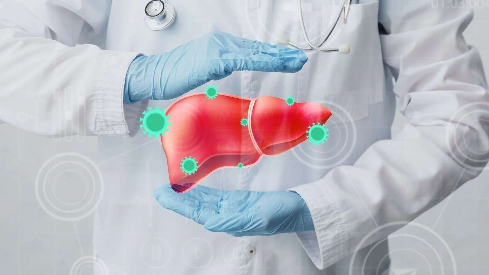
Liver cancer is characterized by malignant tumors that originate in liver tissue and are most commonly referred to as hepatocellular carcinoma. This type of cancer is associated with underlying liver diseases such as cirrhosis and hepatitis B or C infections, and it may not cause symptoms in the early stages.
The most common risk factors for liver cancer include chronic hepatitis infections, long-term alcohol consumption, and fatty liver disease. In addition, exposure to toxins such as aflatoxin can trigger cellular mutations, creating a basis for tumor development.
The diagnostic process is carried out through liver function tests, alpha-fetoprotein (AFP) measurement, and advanced imaging methods. Magnetic resonance imaging (MRI) and computed tomography (CT) are used to evaluate the size and spread of the tumor. A definitive diagnosis is made by biopsy.
The treatment approach varies depending on the stage of the tumor and the patient’s liver function. Surgical resection, liver transplantation, local ablation therapies, and targeted medications are among the main options. Early diagnosis significantly increases treatment success.
| Things You Should Know | Information |
| Definition of Liver Cancer | Tumors that form as a result of the uncontrolled proliferation of liver cells; they may be primary to the liver or metastases from another organ. |
| Primary Liver Cancer | A type of cancer originating from the liver’s own cells (hepatocytes). The most common type is hepatocellular carcinoma. |
| Secondary (Metastatic) Liver Cancer | A type of cancer that begins in another part of the body and spreads to the liver. It may particularly originate from colon, breast, lung, and pancreatic cancers. |
| Most Common Type | Hepatocellular carcinoma (HCC) – develops from the main cells of the liver. |
| Risk Factors | Hepatitis B and C infections, cirrhosis, excessive alcohol consumption, obesity, diabetes, aflatoxin exposure, and genetic diseases (e.g., hemochromatosis). |
| Symptoms of Liver Cancer | Pain in the upper right abdomen, weight loss, loss of appetite, fatigue, jaundice, abdominal swelling (ascites), nausea, and vomiting. |
| Jaundice Symptom | Yellowing of the skin and eyes; indicates impaired liver function. |
| Diagnostic Methods | Physical examination, blood tests (AFP – alpha-fetoprotein), imaging methods (ultrasound, CT, MRI), and biopsy. |
| AFP (Alpha-Fetoprotein) | A tumor marker that increases in liver cancer; helpful in diagnosis. |
| Treatment Options | Surgery (tumor resection), liver transplantation, radiofrequency ablation, chemotherapy, targeted therapies (e.g., sorafenib), immunotherapies. |
| Surgical Treatment | Removal of cancerous tissue in suitable patients; one of the most effective treatment methods. |
| Liver Transplantation | Replacement of the entire liver with a healthy donor liver; may be a permanent solution for some patients. |
| Importance of Early Diagnosis | Treatment success is higher in liver cancers detected at an early stage; treatment options are limited in advanced stages. |
| Prevention Methods | Hepatitis B vaccination, hepatitis C treatment, limiting alcohol consumption, regular health check-ups, and a healthy lifestyle. |
| Follow-up and Monitoring | Regular screening with imaging and AFP testing is recommended for individuals with liver disease. |
What Is Liver Cancer?
Liver cancer is a malignant (cancerous) tumor formed by the uncontrolled proliferation of liver cells. The most common type is hepatocellular carcinoma. It usually develops due to underlying liver diseases such as cirrhosis and hepatitis B or C infections.
What are the types and varieties of primary liver cancer?
When we talk about liver cancer, we are not actually referring to a single disease. Medically, when we say “primary,” we mean that the cancer did not spread from another organ but originated directly from the liver’s own tissue. This distinction is very important because its treatment is completely different from metastases originating from the colon or stomach.
The largest member of this group is Hepatocellular Carcinoma, commonly known as HCC. It develops when hepatocytes, the cells responsible for the liver’s main functions, grow out of control. It ranks among the most common cancers worldwide and is seen slightly more often in men than in women. The second most common type is Cholangiocarcinoma (CC). This type originates from the cells lining the bile ducts inside or outside the liver. Although less common, its behavior and biology are quite different from HCC. It may even present as a special form known as a “Klatskin tumor” at the junction of the bile ducts.
These diseases do not appear overnight. There is usually a damage process that lasts for years. For HCC, suspicious nodules called “dysplastic nodules,” and for bile duct cancers, cellular changes in the ducts (BilIN), are the earliest harbingers on the path to cancer. Our goal is to detect the disease at these early stages or at the very beginning of malignant transformation.
What are the causes and risk factors of liver cancer?
Understanding the causes of the disease is the most important step toward prevention. The risk map we knew in the past has undergone a serious transformation in recent years due to changes in our lifestyle. These risk factors, which vary by geography and habits, must be carefully analyzed.
The main factors that predispose to this disease are:
- Hepatitis B virus
- Hepatitis C virus
- Chronic alcohol consumption
- Non-alcoholic fatty liver disease
- Obesity
- Diabetes
- Aflatoxin exposure
- Smoking
- Genetic predisposition
In previous years, hepatitis B infection was indisputably the number one cause of liver cancer. Although it is still dominant in Asia and Africa, thanks to successful vaccination programs we are seeing a slow but encouraging decline in virus-related cancers. However, the situation is somewhat different in developed countries, where hepatitis C infection remains an important factor.
Today, however, our main concern is a new and rapidly growing enemy: fatty liver disease. Medically known as Non-Alcoholic Fatty Liver Disease (NAFLD), it is a result of the sedentary lifestyle, obesity, and poor dietary habits of the modern era. We are now seeing cancers that develop purely on the basis of metabolic syndrome and excessive fat accumulation, even in patients without viral infections. This clearly shows that liver health is not only about protection from viruses but also about keeping weight and blood sugar under control.
Alcohol consumption alone is a major risk factor, but when combined with viral hepatitis, the situation becomes much more serious. For example, in an individual who is a carrier of hepatitis B or C and also consumes alcohol regularly, the risk increases exponentially rather than additively. A similar “synergistic effect” exists between diabetes and alcohol. In scenarios where diabetes and alcohol coexist, the likelihood of cancer development can reach dramatic levels.
For bile duct cancers (cholangiocarcinoma), the risk factors differ somewhat. Here, conditions that chronically irritate the bile ducts come to the forefront rather than liver cirrhosis. Bile duct cysts, long-standing stones, certain parasitic infections, and rheumatologic diseases such as primary sclerosing cholangitis can create a basis for these cancers.
What are the symptoms of liver cancer?
One of the most challenging aspects of liver cancer is that it is extremely “silent” in its early stages. Because the liver has a very high reserve capacity, it may not send any signals for a long time until a mass reaches a certain size or stretches the organ capsule enough to cause pain. Most of our patients are around 69 years old at the time of diagnosis, which shows how insidiously the process progresses.
When the disease begins to cause symptoms, the most common complaints we encounter are:
- Abdominal pain
- Weight loss
- Loss of appetite
- Fatigue
- Jaundice
- Itching
- Abdominal swelling
- Fever
- Early satiety
When symptoms appear, we usually observe that the underlying cirrhosis has worsened. Yellowing of the eyes and skin, which are signs of progression toward liver failure, abdominal tension due to fluid accumulation (ascites), and persistent itching should raise suspicion of an underlying tumoral formation.
The most common reason for presentation is abdominal pain. Patients usually complain of a dull, constant, and uncomfortable feeling of fullness in the upper right abdomen, beneath the ribs. Sometimes, even after eating a very small amount, a feeling of bloating and fullness occurs. Rarely, we may also encounter systemic symptoms such as low blood sugar, elevated calcium levels, or unexplained changes in blood values due to substances secreted by the tumor. Therefore, regular screening at set intervals is vital for patients in the risk group—those with known cirrhosis or chronic hepatitis—even if they have no complaints.
Is a biopsy necessary during the diagnostic process?
When we suspect liver cancer in a patient, our first stop is imaging methods. Ultrasonography is a good screening tool, but to make a definitive diagnosis, understand the character of the tumor, and plan treatment, we need much more detailed maps. At this point, multiphasic Computed Tomography (CT) or Magnetic Resonance Imaging (MRI) comes into play. These modalities show us the vascular pattern of the tumor, which is the most important fingerprint distinguishing liver cancer from other masses. In addition, tumor markers measured in the blood, such as AFP and PIVKA-II, provide important clues supporting our diagnosis.
So, is it always necessary to take a tissue sample (biopsy)? This is where Interventional Radiology expertise makes a difference. In classical medical practice, biopsy is usually performed by inserting a long needle through the skin. However, since the majority of liver cancer patients have cirrhosis, their coagulation systems may be impaired or they may have fluid accumulation in the abdomen. This can make a conventional needle biopsy a high-risk, dangerous procedure due to bleeding.
To eliminate this risk, we use the Transjugular Liver Biopsy method. This is an extremely elegant and safe technique. We enter through a vein in the patient’s neck (the jugular vein) with a very thin, flexible catheter. Without making any incisions, we advance through the vessels into the liver and take a tissue sample through the vessel wall. Since we do not puncture the outer capsule of the liver, the risk of bleeding into the abdominal cavity is almost zero. This method is a life-saving alternative for patients with coagulation disorders or ascites. Thanks to these possibilities of minimally invasive medicine, we can safely establish a diagnosis even in the highest-risk patients.
What is BCLC staging and treatment planning?
After establishing the diagnosis, the most critical step is determining the stage of the disease. In liver cancer, staging is not done solely based on tumor size. How well the liver is functioning and the patient’s overall performance status are also included in the equation. The Barcelona Clinic Liver Cancer (BCLC) system, which is accepted worldwide, serves as a compass that guides us toward the most appropriate treatment path for our patient.
This system basically divides patients into three main groups: very early/early stage, intermediate stage, and advanced stage. In early-stage patients (BCLC 0/A), our goal is to completely eliminate the disease—that is, to achieve a cure. In the intermediate stage (BCLC B), tumors are usually multiple but without vascular invasion; here, our aim is to control the disease and prolong survival. In the advanced stage, the disease is more widespread, and systemic treatments come to the forefront.
However, this system is not made up of rigid rules. Especially the group we call the intermediate stage is very heterogeneous. In our medical approach, there is flexibility known as “treatment stage migration.” That is, instead of persisting with a treatment that does not yield a response, we can move to a more effective next-line therapy or a different method before liver function deteriorates. Personalized treatment planning is the key to success.
How is tumor ablation performed?
If the tumor is detected at an early stage, it is possible to completely eliminate the disease without the need for surgical intervention, using treatments performed through a needle puncture. The general name for these procedures is Thermal Ablation, meaning the destruction of the tumor with heat.
The principle is quite simple: Under the guidance of imaging devices (ultrasound or CT), we enter through the skin with a special needle and position it directly in the heart of the tumor. Then, by delivering energy through the tip of this needle, we heat the tumor tissue and effectively “cook” it, causing necrosis.
We have two main technologies for this procedure:
- Radiofrequency Ablation (RFA)
- Microwave Ablation (MWA)
Radiofrequency Ablation (RFA) is a reliable method that has been used for many years. It generates heat using electrical current. However, it has some limitations. If the tumor is located right next to a large vessel, the blood flowing through the vessel can carry away the generated heat. This situation, known as the “heat-sink effect,” similar to continuously pouring cold water onto a hot pan, can prevent adequate tumor destruction. In addition, RFA may lose effectiveness in tumors larger than 3 centimeters.
This is where Microwave Ablation (MWA) comes into play. This technology uses electromagnetic waves, similar to the operating principle of microwave ovens, to vibrate water molecules in the tissue and generate very rapid, very high temperatures. The greatest advantage of MWA is that it is much less affected by the cooling effect of nearby vessels. It can also ablate a larger area in a shorter time. It has been shown to be effective even in tumors up to 7–8 centimeters. Moreover, it does not pose a risk of electrical burns. As interventional radiologists, due to these technical advantages, we now more frequently prefer MWA, especially for tumors larger than 3 cm or located near vessels. These procedures are usually performed on a day-case basis; the patient can go home the same day, and post-procedural pain is minimal compared to open surgery.
What is TACE treatment and who is it applied to?
If the number of tumors in the liver is high or their size is too large for ablation, we change our strategy. Instead of targeting tumors one by one, we aim to cut off their main blood supply. This method is called Transarterial Chemoembolization, or TACE for short. It is considered the standard treatment worldwide, especially for intermediate-stage (BCLC B) patients.
The TACE procedure is performed in the angiography unit while the patient is awake. We usually enter through an artery in the groin area. Using very thin, hair-like “microcatheters,” we advance into the liver and then directly into the vessel feeding the tumor. Once we reach the target, we do two things:
- We deliver a high concentration of chemotherapy drug directly into the tumor bed.
- We cut off blood flow by using vessel-occluding particles.
This procedure constitutes a two-pronged siege against the tumor. On one hand, the tumor is exposed to a very high dose of medication; on the other, its blood supply is cut off, leaving it deprived of oxygen and nutrients. This starvation strategy causes the tumor to shrink or die. Because the vessel is occluded, the drug becomes trapped within the tumor and leaks very little into the rest of the body; thus, classic chemotherapy side effects such as hair loss are seen at minimal levels. After the procedure, patients may experience temporary fatigue, nausea, or pain known as “post-embolization syndrome,” but this condition is easily managed with medications and usually resolves within a week.
What is treatment with smart beads (TARE)?
In recent years, with advances in technology, a very powerful alternative to TACE has emerged: Transarterial Radioembolization (TARE), also known as Radioembolization. You may also hear this method referred to among the public as “pinpoint radiation” or “atomic therapy.”
In this treatment, instead of blocking the vessels, we deliver millions of microscopic glass or resin spheres loaded with a radioactive substance called Yttrium-90 (Y90) into them. Again, we enter via the groin using angiographic techniques, locate the vessel feeding the tumor, and release these radioactive beads there. The beads lodge in the capillaries within the tumor and begin to emit very high doses of radiation from the inside out.
The most important difference between TARE and TACE is that TARE does not completely occlude the vessel. Blood flow continues, which is necessary for the effectiveness of radiation (oxygen enhances the power of radiation). As a result, patients usually experience less pain after the procedure and have shorter hospital stays. Scientific studies have shown that TARE is particularly successful in prolonging survival, has a strong tumor-shrinking effect, and offers a better quality of life compared to TACE. It is a powerful weapon that can be safely used even in challenging tumors that have spread to the main portal vein.
What is bridging to liver transplantation?
The definitive solution for liver cancer, if possible, is liver transplantation. However, finding a suitable organ and waiting for one’s turn on the transplant list can take a long time. During this waiting period, it is crucial that the tumor does not grow and eliminate the chance of transplantation. This is where Interventional Radiology methods serve as a “life-saving bridge.”
This is called Bridging therapy. While the patient is on the transplant list, we suppress the tumor by applying TACE, TARE, or ablation, prevent its growth, and ensure that the patient remains eligible on the list.
There is also a concept called Downstaging. Sometimes, when a patient first presents, the tumor is too large to meet transplantation criteria (Milan criteria). In these patients, especially by applying high-dose radioembolization (TARE), we aim to shrink the tumor. If the tumor is reduced to the desired size, the patient may regain eligibility for transplantation. Data show that a significant proportion of patients who were initially not surgical candidates can become transplantable through these methods.
Frequently Asked Questions
What is liver cancer and how does it develop in the body?
What are the earliest symptoms of liver cancer that patients should not ignore?
What causes liver cancer and which risk factors are most significant?
Can liver cancer develop without symptoms in the early stages?
How is liver cancer diagnosed when symptoms are suspected?
What treatment options are available for patients with liver cancer?
When is surgery recommended for the treatment of liver cancer?
What complications can occur if liver cancer is left untreated?
Can lifestyle changes help reduce the risk of developing liver cancer?
What is the prognosis for patients diagnosed with liver cancer?

Prof. Dr. Özgür Kılıçkesmez is one of Türkiye’s leading interventional radiology and neuroradiology specialists, focusing on minimally invasive endovascular treatments. His primary areas of expertise include aneurysm and stroke treatments, embolization procedures, non-surgical treatments of thyroid and parathyroid diseases, interventional oncology, and peripheral vascular interventions. He currently serves as the Head of the Interventional Radiology and Neuroradiology Department at Istanbul Memorial Göztepe Hospital.
Graduating from Istanbul University Cerrahpaşa Faculty of Medicine in 1997, Prof. Dr. Kılıçkesmez received advanced training in interventional radiology and oncology as a Clinical Fellow at London Guy’s & St Thomas’ and King’s College Hospital between 2012 and 2013. He earned the title of Professor in 2020 and, in the same year, became the Founding Interventional Radiologist at SBÜ Çam and Sakura City Hospital. He also holds the EBIR (European Board of Interventional Radiology) certification.
Prof. Dr. Kılıçkesmez has 94 international and 40 national publications and over 2500 citations. He serves as a reviewer for prestigious journals such as the American Journal of Roentgenology (AJR) and Diagnostic & Interventional Radiology. With his clinical and academic expertise, he is a nationally and internationally recognized authority in stroke treatment, brain aneurysm, embolization, tumor ablation, and minimally invasive treatments of thyroid/parathyroid diseases.







Vaka Örnekleri