


Calcification is a condition that occurs when calcium salts accumulate abnormally in body tissues. It is most commonly seen in the breast, vascular structures, kidneys, and soft tissues. It appears as lime-like densities on imaging methods and in some cases may indicate underlying diseases. Not every calcification has to be pathological; some are age-related, degenerative, or incidental benign findings.
Causes of calcium accumulation in tissues include aging, chronic inflammation, tissue damage, and metabolic disorders. Microcalcifications, especially those seen in breast tissue, are detected by mammography and require clinical evaluation to differentiate between benign and malignant processes.
Calcification diagnosis methods are based on radiological imaging techniques. Methods such as mammography, ultrasonography, computed tomography, and magnetic resonance imaging are used to determine the location, size, and distribution of calcium deposits. When necessary, histopathological examination is performed with biopsy.
Calcification treatment is planned according to the underlying cause. Benign and asymptomatic calcifications usually require only regular follow-up. In cases associated with infection, tumors, or metabolic diseases, medical treatment, surgical intervention, or targeted treatment approaches may be applied.
| Things You Should Know | Information |
| Definition of Calcification | Calcification is the condition in which calcium accumulates abnormally in body tissues. This accumulation may occur in soft tissues or organs and usually leads to hardening or calcified formations. |
| Causes of Formation | Trauma, infections, inflammation, certain metabolic diseases (for example hypercalcemia), vascular diseases, aging, some tumors, and genetic factors may cause calcification. |
| Types | 1. Dystrophic Calcification: Accumulates in damaged tissue at normal calcium levels. 2. Metastatic Calcification: Calcium accumulation in normal tissues due to high calcium levels. |
| Most Common Areas | Vessels (in atherosclerotic plaques), heart valves, lungs, kidneys, breast tissue, tendons, and muscles. |
| Signs and Symptoms | It varies depending on the location of the calcification. In some cases, it causes no symptoms, while in others it may cause pain, limited mobility, loss of function, or organ failure. |
| Diagnostic Methods | – X-ray – Ultrasonography – CT (Computed Tomography) – MRI (Magnetic Resonance Imaging) – Blood tests (especially calcium and phosphate levels) – Biopsy (when necessary) |
| Differential Diagnosis | Calcification may be confused with tumors, infections, cysts, or foreign bodies. Differential diagnosis is made through imaging and histopathological examination. |
| Treatment Methods | – Cause-oriented treatment (for example treatment of hypercalcemia) – Surgical intervention (in cases of significant mass or functional impairment) – Physical therapy (especially in musculoskeletal involvement) – Medication (anti-inflammatory, pain relievers, etc.) |
| Prevention Methods | – Control of underlying diseases – Regulation of calcium and phosphate metabolism – Regular health check-ups |
| Follow-up and Monitoring | Since calcification may progress, it should be monitored with imaging and laboratory tests at regular intervals. Careful monitoring is especially necessary in individuals with systemic diseases. |
What Is Calcification and Why Does It Occur in Our Body?
The process of calcification development can actually be considered as a kind of defense or isolation mechanism of the body. No healthy, intact, and well-oxygenated soft tissue normally allows calcium accumulation under normal conditions. Cell membranes have very strict security gates that prevent calcium from entering the tissue uncontrollably. However, when a tissue is exposed to continuous stress, becomes damaged, or begins to lose its vitality for any reason, these security gates collapse.
With cell death or prolonged inflammation in tissues, an acidic environment and harmful molecules called free radicals accumulate in that area. This chemical chaos leads to the rapid attraction of calcium from the blood into the damaged tissue. When the body realizes that it cannot repair the damaged area that has lost its former elasticity, it tries to freeze, isolate, and prevent the problem from spreading further by essentially covering it with lime. These mineral deposits, which initially begin at a microscopic level, can turn into structures that are visible to the eye, can be felt when touched, and even appear as bright and hard as bone on X-ray images over months or years.
Cellular factors that accelerate the process are:
- Inflammation
- Lack of oxygen
- Acidosis
- Cell death
With the combination of these factors, cartilage-like cells proliferate within the soft tissue and secrete calcium crystals, pushing the process into an irreversible path.
What Are the Differences Between Dystrophic and Metastatic Calcification?
To understand mineral accumulations occurring in the body and to establish the correct treatment strategy, dividing the source of this issue into two main categories is of vital importance for the medical field. This distinction shows whether the problem is localized to a specific area or affects the entire body system.
In the mechanism of dystrophic calcification, when the patient’s blood tests are examined, calcium and phosphorus levels are completely within normal limits. The problem is not in the bloodstream or the amount of minerals, but in the tissue itself where calcification occurs. Fat necrosis formed after a breast surgery, calcifications in shoulder tendons that have been strained and worn over years, or calcified plaques in vessel walls beginning to clog due to cholesterol all fall into this group. Since the tissue is diseased, it attracts calcium.
In metastatic calcification, the situation is exactly the opposite. The calcified tissues are actually completely healthy, but the calcium balance in the blood is so disrupted and the levels are so high that the mineral inevitably overflows into soft tissues and begins to deposit. This is like the gates of a dam collapsing and water flooding valleys where it should never be.
Diseases leading to the metastatic process are:
- Hyperparathyroidism
- Sarcoidosis
- Kidney failure
- Hypercalcemia
Without correcting these systemic diseases, it is medically very difficult to stop or reverse calcification in tissues.
How Do Lifestyle and Genetic Factors Affect the Formation of Calcification?
Calcification of tissues in our body is not a random misfortune; our genetic heritage and daily habits maintained over the years weave this process step by step. Especially in calcifications seen in brain tissue and the vascular system, the individual’s life history is the most determining factor. Certain hereditary genetic mutations can predispose to calcium accumulation in deep brain centers that control movement, even in the absence of any metabolic disease.
When it comes to lifestyle, the picture is much clearer. The human body responds to external toxins and poor nutrition by initiating a chronic inflammatory process in the vessel walls. When toxic substances in tobacco products mix with the blood, they literally sandpaper the inner surface of the vessels. In uncontrolled diabetes, sugar crystals that remain constantly high in the blood damage the vessels. Over time, this damage leads to fat accumulation within the vessels and subsequently to calcification and hardening of these deposits.
Lifestyle factors that accelerate calcification are:
- Smoking
- Diabetes
- Hypertension
- Physical inactivity
When these negative factors come together, the elasticity of the vessel wall is completely lost and healthy blood flow is seriously endangered.
Which Imaging Methods Are Used in the Diagnosis of Calcification?
Determining the location, extent, and hardness of calcifications in the body is the most critical step for the success of medical intervention. Interventional radiologists use highly advanced imaging technologies to map the anatomy of calcification. Since calcium tends to block X-rays due to its structure, it appears very clearly in many imaging methods.
Digital mammography has a unique place in the examination of breast tissue. It can detect microcalcifications even smaller than one millimeter. These very small mineral deposits are the first whispers of cellular activity starting in the milk ducts of the breast. Computed tomography is at the peak in detecting the three-dimensional structure of calcifications. By providing a mathematical score of the calcium load in coronary arteries, it allows us to calculate the risk of heart attack. Ultrasonography works on the principle that sound waves hit the hard calcified layer and reflect back, leaving a dark acoustic shadow behind; it provides real-time imaging especially in superficial muscle and joint calcifications and guides needle procedures.
The main devices used are:
- Mammography
- Tomography
- Ultrasonography
- X-ray
These devices are our greatest assistants in determining whether calcification is a benign sign of aging or an indicator of a serious disease.
Do Calcification Findings in the Breast Carry a Cancer Risk?
Reading the phrase “calcification observed in the breast” in a report after routine screening can be a frightening experience for many women. However, not every mineral accumulation in the breast means cancer. In fact, the vast majority of these calcifications are completely harmless and simply reflect the body’s natural aging process, an old trauma, or innocent changes remaining from the breastfeeding period. Radiology specialists classify these findings according to their shapes, sizes, and distribution using an international standard called the BI-RADS system.
Calcifications considered benign and harmless usually appear more regular and larger. Those caused by vascular hardening appear as long thin lines, while those formed within old benign masses look like popcorn. Calcium settled within simple cysts appears in a teacup shape. Such formations do not pose any danger and do not require biopsy. However, some forms may indicate that cells are dividing abnormally and that the process of becoming cancerous has begun at a very early stage.
Calcification features considered risky are:
- Amorphous
- Pleomorphic
- Clustered
- Branching
When calcifications resembling an indistinct cloud of dust, gathered in completely different sizes and shapes, or filling the milk ducts like a broken line are detected, cellular examination becomes mandatory.
How Is Non-Surgical Biopsy Performed in Suspicious Breast Calcifications?
In the past, when a suspicious cluster of calcification was detected in the breast, it was necessary to take the patient to surgery under general anesthesia, make a large incision in the breast, and surgically remove that tissue to understand what it was. Today, with the development of interventional radiology, these procedures can be performed with extremely comfortable, incision-free, and non-surgical methods. Vacuum-assisted biopsy technology is one of the biggest revolutions in this field.
If the calcifications are very small and cannot be seen with ultrasound, special stereotactic systems integrated into the mammography device are used. After the breast is slightly compressed, the device takes images from two different angles and calculates the depth of the calcification within the breast with millimetric precision using a three-dimensional coordinate system. The procedure is performed while the patient is fully awake, with only local anesthesia applied to the breast skin with a thin needle.
The stages of the vacuum biopsy procedure are:
- Coordination
- Anesthesia
- Sampling
- Marking
Only a tiny hole of a few millimeters is opened in the skin. A special needle containing a rotating blade and vacuum mechanism is placed directly at the exact center of the calcifications. When the device is activated, the vacuum power pulls the tissue into the needle, and the blade system removes multiple qualified tissue samples without the patient feeling anything. At the end of the procedure, a small marker clip is left in that area through the same needle so that the location can be easily found in the future. Patients return to their normal lives on the same day without any deformity.
How Are Calcification Diseases That Cause Vascular Occlusion Treated?
Serious health problems such as heart attack and stroke are generally based on vascular occlusions. In standard vascular occlusions, the vessel is entered with closed methods, a deflated balloon is inflated at the blockage site to open the vessel, and then a stent is placed to secure the area. However, when the plaques accumulated in the vessel wall become densely calcified and harden like marble or stone over time, these standard procedures do not work. Trying to expand a hardened vessel with a balloon may cause the vessel to rupture, and even if a stent is placed, it cannot crush the calcified area and cannot open it.
At this point, highly advanced intravascular intervention technologies specific to calcification come into play. The most innovative of these is intravascular lithotripsy, a shock wave therapy that actually works with the logic of kidney stone breaking. A special balloon is placed inside the vessel and inflated at low pressure. The mechanism inside the balloon emits very strong but tissue-safe sound waves outward. These waves focus only on the calcified area and create microscopic cracks within the calcified plaque.
Other advanced technologies used are:
- Lithotripsy
- Rotablator
- Atherectomy
- Laser
In cases where calcification is too thick for shock waves, miniature drill systems with diamond tips rotating hundreds of thousands of times per minute are used. These devices sand the calcified layer into microscopic particles and open the vessel by shaving it. Thanks to all these procedures, even the most difficult vessels can be safely treated without the need for open-heart surgery.
How Does Calcification Causing Severe Shoulder Pain (Calcific Tendinitis) Heal?
Calcifications in the body can sometimes settle directly into the muscles and tendons around the joints. The condition that occurs with the accumulation of calcium crystals in the rotator cuff tendons that move the shoulder joint is called calcific tendinitis. This disease creates such severe pain that patients may wake up crying at night and may become unable to lift their arms even a few centimeters. The calcium within the tendon is sometimes dry like chalk powder and sometimes a thick fluid with the consistency of toothpaste.
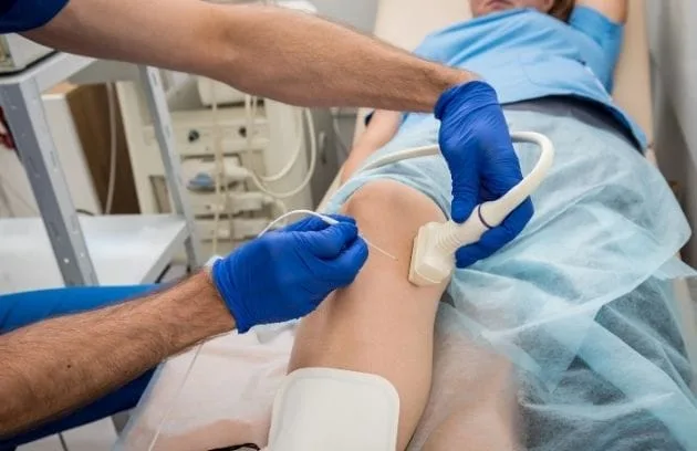
In the past, patients were taken to surgery for this condition, the shoulder was opened, or the tendon was entered with closed camera methods and those stones were scraped out. Today, this condition can be resolved with a very elegant and fast needle treatment called barbotage performed under ultrasound guidance. The interventional radiologist identifies the exact center of the calcium deposit with a high-resolution ultrasound device. After numbing the shoulder skin, a needle is inserted directly into the calcification while watching the ultrasound screen.
The benefits of the procedure for the patient are:
- Speed
- Continuity
- No anesthesia
- Painlessness
Physiological saline is injected through the needle, breaking up and dissolving the toothpaste-like calcification, and then it is aspirated back into the syringe and removed. This washing process continues until the calcification is largely cleared. The procedure takes about fifteen minutes, and since the pressure caused by calcium is immediately relieved, the patient can move their arm much more comfortably even when getting up from the stretcher.
What Are the Advantages of Interventional Radiology Methods Compared to Calcification Surgery?
At the point modern medicine has reached, our goal is to heal the patient while causing the least possible harm, preserving bodily integrity, and ensuring the fastest return to normal life. Needle-hole treatment methods applied in calcification-related health problems offer such significant and clear advantages that they can outcompete traditional open surgeries. First of all, the necessity of general anesthesia, which patients fear most, is eliminated. Patients undergo treatment comfortably while fully conscious, with only the small treatment area being numbed.
In addition, minimal tissue damage creates a huge difference. Making an incision with a scalpel to remove a tumor or calcification means cutting or separating healthy muscles, nerves, and vessels in that area as well. In image-guided procedures, since the target point is visible on the screen, the problem is reached directly with thin needles or intravascular catheters without damaging any healthy tissue.
The main advantages of interventional procedures are:
- Safety
- Comfort
- Aesthetics
- Speed
While patients have to stay in the hospital for days after traditional surgery and endure weeks of physical therapy, these minimally invasive procedures are performed on an outpatient basis. A patient who comes to the clinic in the morning can receive treatment and rest at home in the afternoon without any permanent stitches or incision scars on the body.
How Are Calcification Problems in Different Organs Managed?
Calcification is not just a localized problem seen in the breast, shoulder, or vessels; it has a wide spectrum that can threaten the structure and function of the most vital organs of the body. In particular, the thickening and calcification of the pericardium surrounding the heart over the years prevents the heart from expanding comfortably and filling with blood with each beat. This condition traps the heart in an armored cage, leading to severe signs of heart failure. Treatment of these patients may require highly detailed pericardial stripping surgeries after tomography mapping.
The kidneys are the body’s filters and one of the main centers of calcium metabolism. Increased calcium levels in urine or changes in urine acidity can directly lead to widespread calcification within the kidney tissue itself. If calcium stones accumulated in the kidney channels block urine flow, the kidney begins to deteriorate.
Other organs and structures that may be affected are:
- Heart
- Pericardium
- Kidneys
- Brain
In such cases, stones are broken with high-frequency sound waves sent from outside, or urine pathways are safely drained by entering the kidney through the skin with a needle. Calcium accumulations in brain tissue, especially in movement centers due to genetic or metabolic causes, are generally irreversible; at this point, the main goal is to control the underlying hormonal disorder or mineral imbalance with medication.
Can the Progression of Calcification Be Stopped or Prevented?
Although it is not entirely possible to stop tissue calcification before it even begins, since it is a natural result of aging and cellular wear, slowing down this process and preventing it from reaching life-threatening levels is definitely under the patient’s control. Dystrophic calcifications, especially those associated with vascular hardening and tissue degeneration, are directly fed by our lifestyle. When we remove factors that fuel inflammation in our body from our lives, the progression of calcification comes to a halt.
It should not be forgotten that nutrition directly affects calcium accumulation. Calcium or vitamin D supplements taken unconsciously, in high doses, and without testing, based on hearsay information, accumulate directly in the kidneys and vessel walls instead of going to the bones. Keeping blood pressure at normal levels reduces physical pressure and damage on the vessel wall, eliminating the basis of calcification.
The main strategies to be implemented are:
- Exercise
- Diet
- Control
- Awareness
Frequently Asked Questions
What Is Calcification and why does it occur in the body?
Calcification is the accumulation of calcium deposits within tissues, blood vessels, or organs. It can develop as a result of aging, chronic inflammation, tissue injury, metabolic disorders, or certain underlying medical conditions.
What Is Calcification and can it affect different parts of the body?
Calcification can occur in many areas, including arteries, heart valves, breasts, kidneys, joints, and soft tissues. The impact depends on the location, size, and extent of the calcium deposits.
What symptoms can develop when calcification becomes significant?
Many calcifications cause no symptoms, especially in early stages. However, larger or strategically located deposits may lead to pain, stiffness, reduced organ function, circulation problems, or other condition-specific symptoms.
How Is Calcification Diagnosed during routine medical evaluation?
Calcification is often discovered incidentally during imaging studies performed for other reasons. Physicians may also investigate calcification when patients present with unexplained symptoms or risk factors for chronic disease.
How Is Calcification Diagnosed using imaging techniques?
Doctors commonly use X-rays, CT scans, ultrasound, mammography, or MRI studies to identify calcium deposits. The choice of imaging depends on the affected body region and the suspected underlying condition.
Can blood tests help diagnose the cause of calcification?
Blood tests may evaluate calcium, phosphate, vitamin D, kidney function, and hormonal abnormalities. While they do not directly visualize calcification, they can help identify contributing metabolic or systemic disorders.
What risk factors increase the likelihood of developing calcification?
Common risk factors include aging, diabetes, chronic kidney disease, cardiovascular disease, smoking, chronic inflammation, elevated calcium metabolism disorders, and long-standing tissue injury or degeneration.
How Is Calcification Treated when symptoms are present?
Treatment depends on the location and underlying cause. Options may include medications, lifestyle modifications, management of associated diseases, minimally invasive procedures, or surgery in selected cases.
Can lifestyle changes help prevent the progression of calcification?
Maintaining a healthy diet, controlling blood pressure, avoiding tobacco use, managing diabetes, and staying physically active may help reduce the risk of progression, particularly in vascular calcification.
Can Calcification be reversed after it has developed?
Some forms of calcification can be stabilized or managed, but complete reversal is often difficult. Early detection and treatment of underlying conditions may help slow progression and reduce complications.

Prof. Dr. Özgür Kılıçkesmez is one of Türkiye’s leading interventional radiology and neuroradiology specialists, focusing on minimally invasive endovascular treatments. His primary areas of expertise include aneurysm and stroke treatments, embolization procedures, non-surgical treatments of thyroid and parathyroid diseases, interventional oncology, and peripheral vascular interventions. He currently serves as the Head of the Interventional Radiology and Neuroradiology Department at Istanbul Memorial Göztepe Hospital.
Graduating from Istanbul University Cerrahpaşa Faculty of Medicine in 1997, Prof. Dr. Kılıçkesmez received advanced training in interventional radiology and oncology as a Clinical Fellow at London Guy’s & St Thomas’ and King’s College Hospital between 2012 and 2013. He earned the title of Professor in 2020 and, in the same year, became the Founding Interventional Radiologist at SBÜ Çam and Sakura City Hospital. He also holds the EBIR (European Board of Interventional Radiology) certification.
Prof. Dr. Kılıçkesmez has 94 international and 40 national publications and over 2500 citations. He serves as a reviewer for prestigious journals such as the American Journal of Roentgenology (AJR) and Diagnostic & Interventional Radiology. With his clinical and academic expertise, he is a nationally and internationally recognized authority in stroke treatment, brain aneurysm, embolization, tumor ablation, and minimally invasive treatments of thyroid/parathyroid diseases.





Vaka Örnekleri