


A liver tumor is an abnormal growth of cells within liver tissue that may be benign or malignant, arising from hepatocytes or other cellular components. These lesions vary in size, structure, and clinical significance, requiring accurate diagnosis to determine appropriate medical management and prognosis.
Liver tumor types and classifications include benign forms such as hemangiomas and adenomas, as well as malignant variants like hepatocellular carcinoma and metastatic lesions. Each type demonstrates distinct pathological features, growth patterns, and potential complications affecting liver function and systemic health.
Symptoms of liver tumors and clinical presentation often involve abdominal discomfort, unexplained weight loss, jaundice, and fatigue in advanced stages. Early-stage tumors may remain asymptomatic, making imaging techniques and laboratory evaluations essential for timely detection and intervention.
Diagnosis and treatment of liver tumors depend on tumor type, size, location, and patient health status. Imaging modalities such as ultrasound, CT, and MRI, combined with biopsy, guide management strategies including surgical resection, ablation therapies, chemotherapy, or targeted treatments.
| What You Need to Know | Information |
| Liver Tumors | Abnormal cell growths occurring in the liver; can be benign or malignant. |
| Benign Tumors | Usually asymptomatic and discovered incidentally. Most common types are hemangioma, hepatic adenoma, and focal nodular hyperplasia (FNH). |
| Malignant Tumors | Malignant liver tumors can be primary (originating in the liver) or metastatic (spreading from other organs). |
| Primary Malignant Tumors | The most common is hepatocellular carcinoma (HCC). Others include cholangiocarcinoma (bile duct cancer), hepatoblastoma (seen in children), and angiosarcoma. |
| Metastatic Tumors | The most common cancers that metastasize to the liver originate from the colon, breast, lungs, and pancreas. Most malignant tumors in the liver are of metastatic origin. |
| Hemangioma | The most common benign liver tumor. Usually small, asymptomatic, and does not require treatment. |
| Hepatic Adenoma | More common in women who use birth control pills. Larger ones may be surgically removed due to the risk of bleeding or malignant transformation. |
| Focal Nodular Hyperplasia (FNH) | The second most common benign liver lesion. Generally does not require treatment. Diagnosis can be made through imaging. |
| Hepatocellular Carcinoma (HCC) | Develops in the setting of chronic liver disease or cirrhosis. Risk factors include Hepatitis B/C and alcohol use. |
| Cholangiocarcinoma | Originates from the bile ducts. Usually presents late and is difficult to diagnose. Surgery is the main treatment method. |
| Symptoms | Symptoms may include abdominal pain, weight loss, jaundice, a feeling of a mass in the abdomen, and loss of appetite. However, early stages may not show any symptoms. |
| Diagnostic Methods | Ultrasonography, CT (computed tomography), MRI (magnetic resonance imaging), and biopsy are commonly used. |
| Treatment Options | Surgical resection, liver transplantation, radiofrequency ablation, chemotherapy, and targeted therapy options may be applied. |
| Importance of Early Diagnosis | Early diagnosis increases treatment success and life expectancy, especially in malignant tumors like HCC. |
| Follow-up and Monitoring | Patients in the risk group should be followed with regular imaging. Patients with cirrhosis should be monitored every 6 months with ultrasound. |
What Is a Liver Tumor?
A liver tumor refers to benign or malignant cell growths that develop in the liver. Benign tumors generally do not present symptoms, while malignant ones are defined as liver cancer (such as hepatocellular carcinoma). Tumors may be primary (originating in the liver) or metastatic (spreading from another organ). If symptoms occur, they may include abdominal pain, weight loss, and jaundice. Treatment is planned according to the tumor type and its spread.
What Are the Types and Classifications of Liver Tumors?
The first and most crucial step in understanding liver masses is to determine whether the mass is benign or malignant. Benign tumors are generally stationary, do not invade surrounding tissues, and do not spread (metastasize) to other parts of the body. Malignant tumors, on the other hand, are cancerous tissues that grow uncontrollably and can be life-threatening.
Malignant tumors are further classified into two main groups. The first group includes “primary” cancers originating directly from the liver’s own cells. The second group consists of “secondary” (metastatic) tumors, which develop when cancers that begin in other organs—such as the colon, stomach, breast, or lungs—spread to the liver through the bloodstream. This distinction is vital because the treatment strategy for primary liver cancer is completely different from that used for metastases.
The most commonly encountered tumor groups in the liver are:
- Hemangioma
- Focal Nodular Hyperplasia
- Hepatocellular Carcinoma
- Intrahepatic Cholangiocarcinoma
- Colorectal Liver Metastases
- Hepatocellular Adenoma
Can a benign liver tumor be mistaken for cancer?
One of the greatest challenges in liver imaging is that benign formations can sometimes visually mimic cancerous tissues. The most common benign liver tumor is a structure called a “Hemangioma.” This is a cluster of blood vessels, often congenital and usually requiring no treatment. However, in some cases, the radiological appearance of hemangiomas can be so complex that it becomes difficult to distinguish them from hepatocellular carcinoma, the primary cancer of the liver.
Similarly, the second most common benign tumor, “Focal Nodular Hyperplasia” (FNH), is essentially an overgrowth response of liver tissue to a vascular abnormality. FNH is definitely not cancer and does not turn into cancer. However, certain features under the microscope or on MRI images can unfortunately resemble malignant tumors. At this point, a detailed examination process called “differential diagnosis” comes into play. If we mistakenly consider a harmless FNH or hemangioma as cancer and attempt surgery, we put the patient at unnecessary risk. Conversely, if we assume a cancerous tissue is benign and choose to monitor it, the disease may progress. Therefore, from an interventional radiology perspective, it is crucial to use specialized imaging techniques in suspicious cases and, if necessary, obtain a tissue sample with a needle biopsy before initiating treatment.
Benign lesions that may appear confusing on imaging include:
- Hemangioma
- Focal Nodular Hyperplasia
- Hepatic Angiomyolipoma
- Intrahepatic Splenosis
- Inflammatory Pseudotumor
What is Hepatocellular Carcinoma (HCC), the primary liver cancer?
Hepatocellular Carcinoma (HCC) is the most common primary liver cancer in the world and originates from the liver’s main cells. This type of cancer typically does not develop suddenly in a healthy liver. It usually arises as a result of years of chronic damage. When liver cirrhosis develops due to chronic Hepatitis B or C infection, excessive alcohol consumption, or fatty liver disease, genetic errors can occur in the regenerating liver cells, leading to cancer.
HCC is the second leading cause of cancer-related deaths worldwide, highlighting the seriousness of the disease. Internationally accepted staging systems (such as the BCLC staging) are used to determine the stage at diagnosis. This staging evaluates not only tumor size but also how well the liver is functioning and the patient’s overall performance status. In early-stage tumors (under 2–3 cm), the chance for a full cure is very high. In advanced stages where the tumor has spread to blood vessels or enveloped the liver, treatment options become more complex.
The main risk factors involved in the development of HCC are:
- Chronic Hepatitis B infection
- Chronic Hepatitis C infection
- Liver cirrhosis
- Excessive alcohol consumption
- Non-alcoholic fatty liver disease
Why do cancers from other organs metastasize to the liver?
The liver is one of the most highly vascularized organs in the body and is located at a very strategic junction within the circulatory system. Blood from the intestines, stomach, pancreas, and spleen flows into the liver via a large vein called the “Portal Vein” for detoxification and processing. This anatomical structure unfortunately makes the liver a very suitable target for cancer cells from other organs to settle. Especially colon cancers are the most common cancers to metastasize to the liver, due to the direct blood flow path. Medically, this is referred to as “Colorectal Liver Metastasis” (CRLM).
In the past, liver metastasis was considered the final stage of cancer, and few treatment options were available. Today, this perspective has completely changed. If the number of metastases is limited (called oligometastatic disease) and liver function is preserved, we can treat these metastases as if they were primary tumors. Interventional methods to eliminate these lesions can significantly prolong survival and, in some suitable patients, even achieve complete recovery.
What methods are used to diagnose liver tumors?
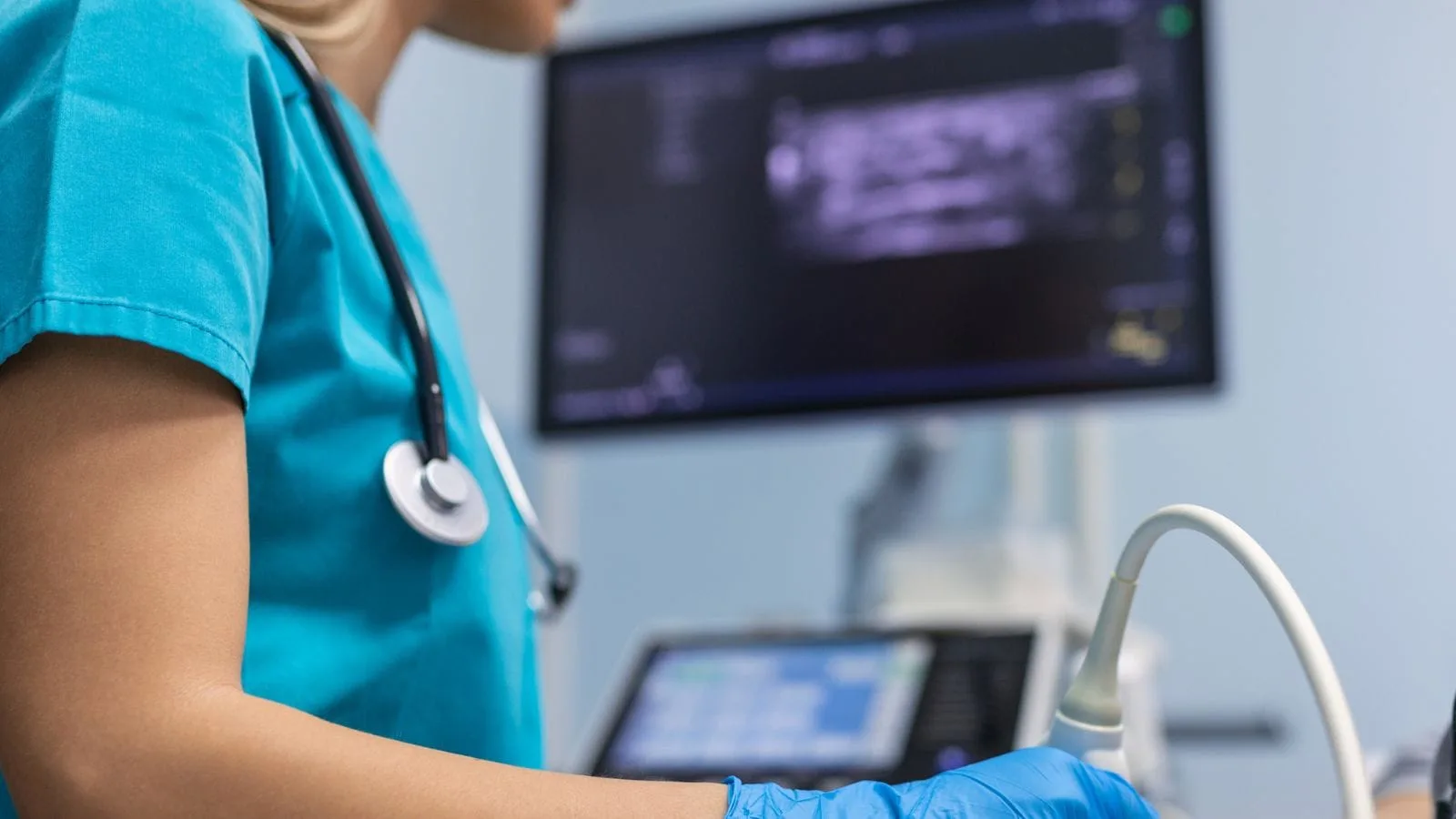
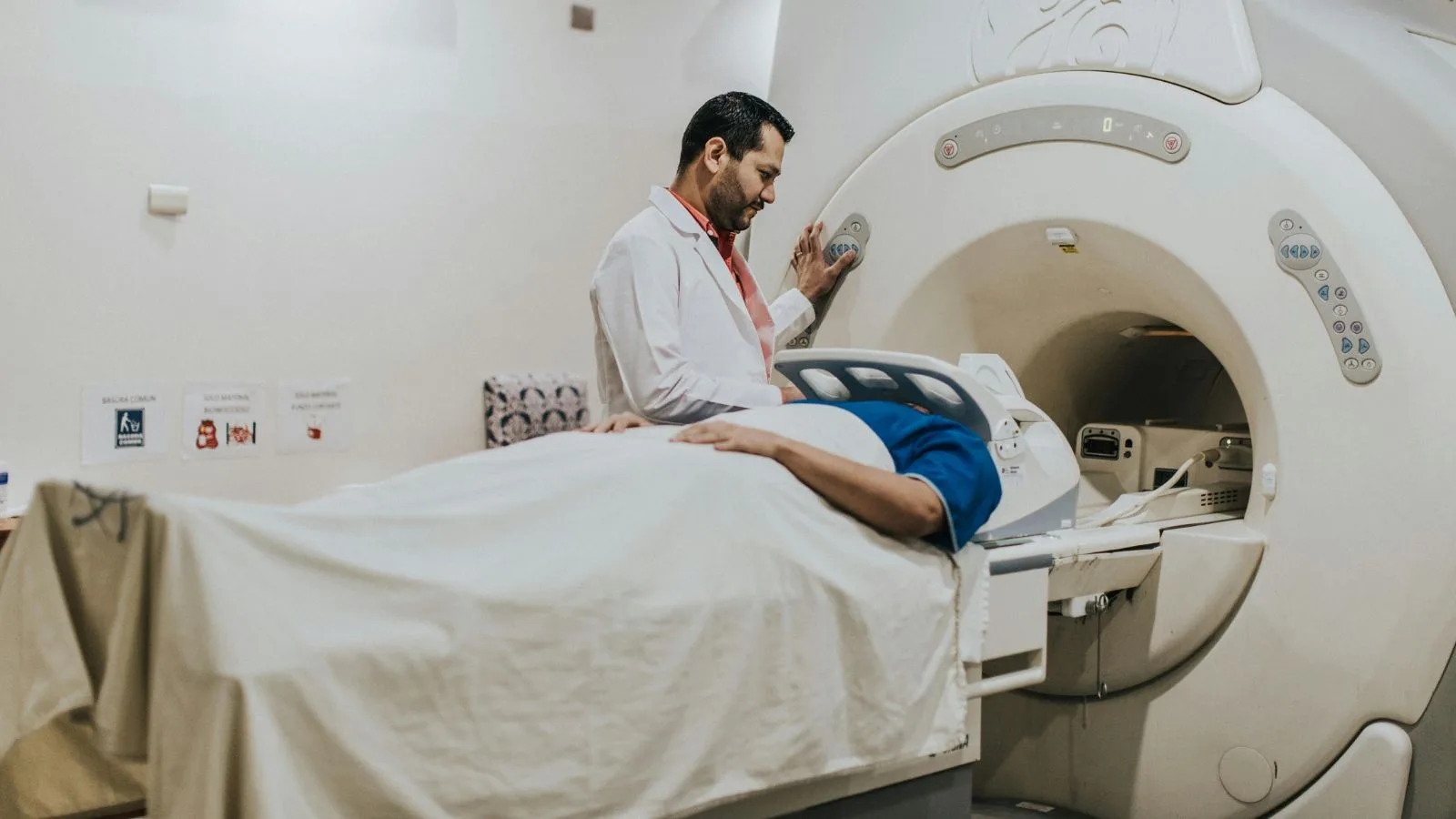
When a liver mass is suspected, the diagnostic process requires detective-like investigation. It usually begins with a review of the patient’s clinical history, hepatitis status, and blood tests. However, the main determinant is high-tech radiological imaging. Ultrasonography is generally used as the first screening method, but “multiphasic” Computed Tomography (CT) or Magnetic Resonance Imaging (MRI) is essential to clarify the nature of the tumor.
“Multiphasic” imaging involves administering contrast material intravenously and taking a series of scans of the liver in different time phases (within seconds). Each tumor has a unique vascular pattern. For example, HCC-type cancers quickly absorb the contrast in the arterial phase and then rapidly wash it out. Hemangiomas, on the other hand, fill with contrast much more slowly, from the edges inward. These characteristic enhancement patterns often allow for diagnosis without biopsy. However, if the imaging is inconclusive, a thin needle biopsy through the skin may be necessary to confirm the diagnosis.
The main diagnostic tools used in this process include:
- Ultrasonography
- Multiphasic Computed Tomography
- Dynamic Magnetic Resonance Imaging
- Percutaneous Needle Biopsy
- Alpha-fetoprotein blood test
What Is Minimally Invasive Treatment Through a Needle Hole (Interventional Radiology)?
Interventional Radiology is a field where medicine merges with technology, involving treatments performed without large surgical incisions—only through needle punctures or via blood vessels. While surgical removal of the tumor (resection) has historically been the most effective method in liver tumors, the majority of patients are not suitable for surgery due to underlying cirrhosis or the location of the tumor. At this point, interventional methods focused directly on the tumor, known as “Locoregional Therapies,” come into play.
The core philosophy of these treatments is to intervene directly at the site of the problem, rather than administering harsh drugs that affect the whole body. Using imaging devices (angiography, ultrasound, CT) as guidance, the tumor is reached with millimeter precision. This approach preserves healthy liver tissue while destroying the cancerous tissue. These methods shorten hospital stays, speed up recovery, and preserve patients’ quality of life. Treatment options vary from burning the tumor (ablation) to blocking its blood supply (embolization).
How Is Tumor Burning (Ablation) Performed?
Ablation therapy is a highly effective method that offers surgical-equivalent outcomes for small liver tumors under 3 cm. The procedure is performed under mild sedation and local anesthesia for patient comfort. The interventional radiologist visualizes the tumor using ultrasound or CT and advances a specially designed needle through the skin directly into the center of the tumor.
Depending on the technology used, Radiofrequency (RFA) or Microwave (MWA) energy is applied, generating high heat at the needle tip. This heat “cooks” the tumor tissue and a small surrounding safety margin, killing the cells instantly. Microwave ablation is a newer technology that can reach higher temperatures more quickly, making it useful for slightly larger tumors or those close to blood vessels. The tumor is completely destroyed and eventually shrinks over time within the body. In oligometastatic colon cancer metastases or early-stage HCC, this method can offer a chance of complete cure while avoiding major surgery.
Advantages of ablation therapy include:
- Short procedure time
- Same-day or one-night hospital stay
- Low complication risk
- Repeatable procedure
- No surgical incision scars
How Does Intra-arterial Chemotherapy (TACE) Destroy the Tumor?
Transarterial Chemoembolization (TACE) is the standard treatment, especially for intermediate-stage liver cancers with multiple tumors or those too large for ablation. The core idea of this method is to cut off the tumor’s blood supply. While normal liver tissue receives most of its blood from the portal vein (coming from the intestines), liver tumors are primarily fed by the hepatic artery. This biological difference forms the basis of the treatment.
During the procedure, a catheter is inserted through the groin vein and guided under angiography to the hepatic artery. From there, much thinner microcatheters are advanced directly into the arteries feeding the tumor. A two-step attack is then carried out: First, a high dose of chemotherapy is delivered directly to the tumor bed; then, the artery is blocked with special particles. As a result, the tumor is subjected to a concentrated dose of toxic drugs and is deprived of oxygen, essentially starving to death. Since the drug is trapped within the tumor, systemic side effects (like hair loss) are significantly reduced compared to traditional chemotherapy.
Why Is Success Higher When TACE and Ablation Are Used Together?
In medicine, combining two different methods can sometimes create a more powerful effect than using either alone. Especially for tumors between 3 and 5 cm, ablation alone may not completely destroy all cells, particularly on the tumor’s outer edge. One reason is the cooling effect of surrounding blood flow, which reduces the heating efficacy.
For tumors of this size, the combination of “TACE + Ablation” has been developed. First, TACE is performed to block blood flow and deliver chemotherapy. Once blood flow is cut off, the tumor loses its cooling effect and becomes more vulnerable to heat. About 2 to 4 weeks after TACE, ablation is applied to burn and destroy the now poorly perfused tumor more effectively. Scientific data shows that this combined approach offers up to a 90% complete response rate for tumors between 3–5 cm and significantly reduces recurrence risk.
What Is Radiation Therapy with Radioactive Particles (TARE / Radioembolization), and Who Is It For?
Transarterial Radioembolization (TARE), commonly known as “Radiation Therapy” or “Atom Therapy,” is one of the most powerful weapons against liver tumors. Similar to the TACE procedure, the method involves accessing the tumor via the bloodstream. However, instead of blocking the vessel, millions of microscopic beads loaded with the radioactive substance Yttrium-90 (Y-90) are delivered.
These radioactive beads lodge in the tumor’s capillaries and begin to emit very high doses of radiation from the inside out, but over very short distances. Because the radiation range is measured in millimeters, the healthy liver tissue next to the tumor remains unharmed. This method is particularly effective for large tumors (up to 8 cm) or cancers that have spread into major vessels like the portal vein. TARE is not just a palliative (symptom-relieving) treatment; it can shrink tumors to prepare them for surgery or even eliminate large masses entirely, with curative potential. It plays a strategic role especially when chemotherapy is insufficient or when preserving liver function is critical.
Indications for TARE therapy include:
- Large-sized HCC tumors
- Patients with portal vein thrombosis
- Liver-limited colon cancer metastases
- Pre-surgical tumor shrinkage
- Chemotherapy-resistant tumors
How Is the Treatment Decision Made, and What Is the Process?
The management of liver tumors is not simple enough for a single physician to decide. A “Multidisciplinary Tumor Board” approach is adopted to fight this complex disease. This board includes interventional radiologists, medical oncologists, liver surgeons, gastroenterologists, and nuclear medicine specialists. Each patient’s case, imaging, and overall health are discussed in detail.
In making the treatment decision, not only the size of the tumor but also the liver’s remaining functional reserve, the patient’s age, and comorbidities are considered. For example, surgical and ablation options may be compared for a small tumor, while for a patient with multiple metastases, the timing of chemotherapy and TARE may be planned. As interventional radiologists, our role in this team is to identify and apply the most advanced, minimally invasive technique that extends the patient’s life with the best possible quality. The most accurate roadmap for our patients can only be drawn in the light of this shared wisdom and scientific guidelines.
Frequently Asked Questions
What is a liver tumor and how does it develop in the liver tissue?
What is a liver tumor and what are the earliest symptoms patients may notice?
What is a liver tumor and can abdominal pain indicate its presence?
What is a liver tumor and why can unexplained weight loss occur?
What is a liver tumor and how is jaundice related to the condition?
What is a liver tumor and who is at increased risk of developing one?
What is a liver tumor and how is it diagnosed by healthcare professionals?
What is a liver tumor and can benign liver tumors cause symptoms?
What is a liver tumor and what treatment options are available?
What is a liver tumor and when should medical attention be sought for symptoms?

Prof. Dr. Özgür Kılıçkesmez is one of Türkiye’s leading interventional radiology and neuroradiology specialists, focusing on minimally invasive endovascular treatments. His primary areas of expertise include aneurysm and stroke treatments, embolization procedures, non-surgical treatments of thyroid and parathyroid diseases, interventional oncology, and peripheral vascular interventions. He currently serves as the Head of the Interventional Radiology and Neuroradiology Department at Istanbul Memorial Göztepe Hospital.
Graduating from Istanbul University Cerrahpaşa Faculty of Medicine in 1997, Prof. Dr. Kılıçkesmez received advanced training in interventional radiology and oncology as a Clinical Fellow at London Guy’s & St Thomas’ and King’s College Hospital between 2012 and 2013. He earned the title of Professor in 2020 and, in the same year, became the Founding Interventional Radiologist at SBÜ Çam and Sakura City Hospital. He also holds the EBIR (European Board of Interventional Radiology) certification.
Prof. Dr. Kılıçkesmez has 94 international and 40 national publications and over 2500 citations. He serves as a reviewer for prestigious journals such as the American Journal of Roentgenology (AJR) and Diagnostic & Interventional Radiology. With his clinical and academic expertise, he is a nationally and internationally recognized authority in stroke treatment, brain aneurysm, embolization, tumor ablation, and minimally invasive treatments of thyroid/parathyroid diseases.







Vaka Örnekleri