Arteriovenous malformations (AVM) are abnormal connections between arteries and veins that disrupt normal blood circulation. They may cause pain, bleeding, or neurological symptoms, and require precise diagnosis and individualized treatment strategies.
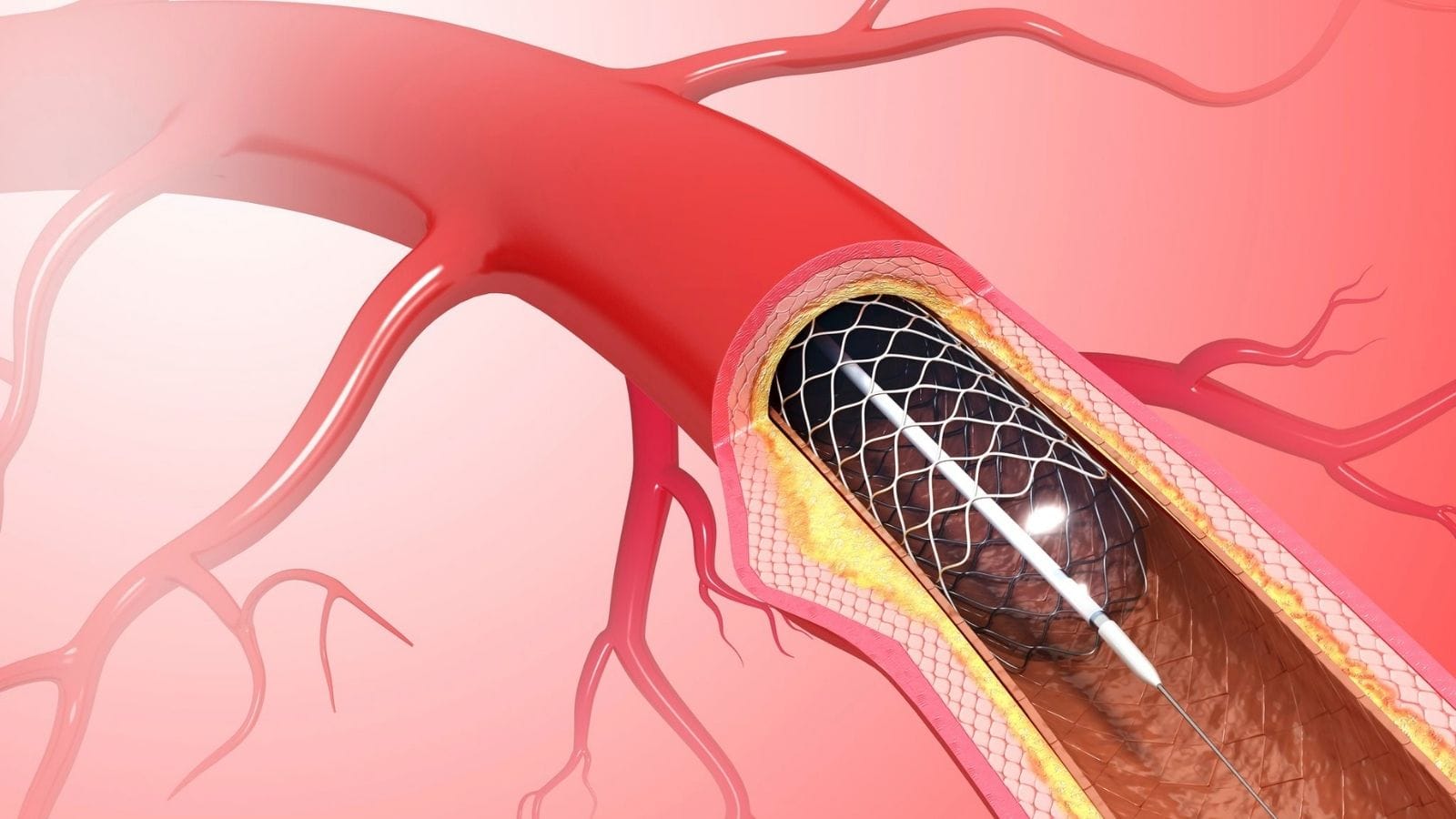
Interventional radiology offers minimally invasive options for AVM treatment. Embolization techniques block abnormal blood flow, shrink malformations, and reduce risks of rupture, often as a standalone therapy or in preparation for surgery.
AVMs can occur in various organs including the brain, lungs, and extremities. Clinical presentation depends on location, with cerebral AVMs posing risks of seizures and hemorrhage, while peripheral AVMs often cause swelling and pain.
Advanced imaging methods such as MRI and angiography are essential in diagnosis and treatment planning. These modalities provide detailed vascular mapping, enabling safe and effective interventional or surgical approaches tailored to each patient.
| Definition | Arteriovenous malformation (AVM) is an abnormal connection between arteries and veins. These connections are congenital and characterized by the lack of a normal capillary bed. They can occur anywhere in the body. |
| Causes | Congenital (genetic) with an unknown exact cause. Genetic factors may play a role. |
| Symptoms | In those located in the brain, symptoms can include headaches, seizures, neurological deficits (vision, speech, or movement disorders), dizziness, tinnitus, bleeding (intracranial bleeding), and loss of consciousness. In extremities, body areas, or musculoskeletal systems, swelling, pain, discoloration, and the presence of a murmur are common findings. Rarely, they may thin the skin and cause bleeding. |
| Diagnostic Methods | MRI and MR angiography, computed tomography (CT) and CT angiography, angiography, Doppler ultrasound |
| Treatment Methods | Microsurgical resection, endovascular embolization, stereotactic radiosurgery (Gamma Knife or CyberKnife) for brain lesions, medication (for symptom management). For peripheral (non-brain) lesions, the main option is endovascular embolization. These lesions often extend into many tissues and cannot be easily removed. |
| Procedure Types | Minimally invasive (endovascular embolization), surgical (microsurgical resection), radiosurgery (stereotactic radiosurgery) |
| Procedure Duration | Varies depending on the treatment method; surgery typically lasts a few hours, while embolization and radiosurgery may be quicker. Endovascular treatment typically leads to a cure in brain lesions, while repeated treatments may be required for those located in the body. |
| Anesthesia Type | Typically general anesthesia, or deep sedoanalgesia |
| Preparation | Detailed neurological evaluation, blood tests, imaging tests, preoperative evaluation |
| Side Effects and Risks | Infection, bleeding, neurological deficits, anesthesia complications, ischemic complications post-embolization, skin wounds |
| Recovery Time | Several weeks to several months post-surgery; faster recovery after endovascular and radiosurgery |
| Follow-up | Regular neurological examinations, follow-up imaging, monitoring for potential complications post-treatment |
| Advantages | Various treatment options, availability of minimally invasive methods, reduction of symptoms and bleeding risk with treatment |
| Alternative Treatments | Symptomatic treatment (antiepileptic drugs), conservative monitoring (regular imaging and clinical follow-up) |

Prof. Dr. Özgür KILIÇKESMEZ
Interventional Radiology / Interventional Neuroradiology
Prof. Dr. Kılıçkesmez holds the Turkish Radiology Competency Certificate, the Turkish Interventional Radiology Competency Certificate, Stroke Treatment Certification, and the European Board of Interventional Radiology (EBIR). In his academic career, he won the Siemens Radiology First Prize in 2008.
Definition and Effects of AVM Disease

- Bleeding brain vascular mass (AVM) embolization treatment
Arteriovenous malformation, or AVM, is an abnormal fusion of blood vessels. This condition is usually a congenital vascular mass that can occur anywhere in the body. When located in critical areas such as the nervous system, it can lead to severe health issues.
AVMs create direct connections between arteries and veins instead of normal capillary beds. These abnormal connections affect blood flow and may lead to various complications:
- Blood is less efficient in delivering oxygen and nutrients to cells,
- Waste products may not be properly eliminated from the body.
- Excessive blood flow can cause swelling and increased warmth in the affected limbs.
The exact formation mechanism of AVM is not fully understood, though genetic factors may play a role. The presence of this structural anomaly can lead to functional disorders in related organs and severe health problems over time. Therefore, when AVM is diagnosed, it must be closely monitored and managed according to its severity.
*We recommend filling out all fields so we can respond in the best possible way.
Symptoms and Signs of Arteriovenous Malformation
Arteriovenous malformations (AVMs) typically occur within the nervous system and often do not show symptoms. However, once symptoms begin, they can vary greatly between individuals.
Brain AVMs, by affecting blood flow in the brain, can lead to general complaints such as headaches. Depending on the location and the brain area affected, additional symptoms may appear:
- Weakness on one side of the body
- Numbness
- Loss of balance
- Speech difficulties
- Dementia
- Intellectual disability in children
- Seizures
Spinal AVMs present a different set of symptoms:
- Back and lower back pain
- Numbness in the legs
- Weakness
- Urinary incontinence
- Paralysis due to sudden bleeding
These symptoms vary depending on the location and severity of the AVM. Without treatment, these symptoms can worsen, leading to serious health problems. Therefore, early diagnosis and intervention are essential.
Body AVMs:
- Thickening of the arms and legs
- Skin discoloration
- Warmth over the lesion, while the surrounding area may be cold.
AVM Disease Treatment Methods

- lung avm image
In arteriovenous malformations, lesions with high risk require special attention. In such cases, alternatives to surgical intervention are preferred. Endovascular embolization is an important method used to reduce the lesion’s risk.
This treatment facilitates the closure of the tangled vessels, minimizing the risk of potential bleeding. On average, two years after treatment, the tangled vessels are fully closed. Furthermore, this method:
- Facilitates closure of the lesion,
- Reduces pre-surgical risks,
- Reduces the likelihood of bleeding.
Endovascular embolization offers a less invasive solution compared to surgery and provides a faster recovery process for patients.

Girişimsel Radyoloji ve Nöroradyoloji Uzmanı Prof. Dr. Özgür Kılıçkesmez, 1997 yılında Cerrahpaşa Tıp Fakültesi’nden mezun oldu. Uzmanlık eğitimini İstanbul Eğitim ve Araştırma Hastanesi’nde tamamladı. Londra’da girişimsel radyoloji ve onkoloji alanında eğitim aldı. İstanbul Çam ve Sakura Şehir Hastanesi’nde girişimsel radyoloji bölümünü kurdu ve 2020 yılında profesör oldu. Çok sayıda uluslararası ödül ve sertifikaya sahip olan Kılıçkesmez’in 150’den fazla bilimsel yayını bulunmakta ve 1500’den fazla atıf almıştır. Halen Medicana Ataköy Hastanesi’nde görev yapmaktadır.


Vaka Örnekleri