


An aneurysm is a localized dilation of a blood vessel wall caused by structural weakness, most commonly occurring in cerebral arteries. A brain aneurysm may remain asymptomatic or rupture, leading to subarachnoid hemorrhage, a life-threatening neurological emergency requiring immediate intervention.
Brain aneurysm symptoms without rupture often include localized headache, visual disturbances, cranial nerve deficits, and orbital pain due to pressure on adjacent neural structures. These manifestations vary depending on aneurysm size, location, and rate of expansion within intracranial circulation.
Ruptured brain aneurysm symptoms typically present with sudden, severe headache described as the worst headache of life, accompanied by nausea, vomiting, neck stiffness, photophobia, and altered consciousness. Rapid neurological deterioration may occur due to increased intracranial pressure and bleeding.
Risk factors for brain aneurysm formation and rupture include hypertension, smoking, genetic predisposition, connective tissue disorders, and advanced age. Early diagnosis through imaging modalities such as CT angiography and timely neurosurgical or endovascular treatment significantly improves clinical outcomes.
| What You Should Know | Information |
| What Is an Aneurysm? | An enlargement of the vessel wall, usually in the form of a balloon. It is most commonly seen in arteries. |
| What Is a Brain Aneurysm? | An aneurysm that forms in the arteries of the brain. When it ruptures, bleeding into the brain (subarachnoid hemorrhage) occurs. |
| Prevalence | Seen in approximately 1–2% of the population. Not every aneurysm results in bleeding, but it carries risk. |
| Most Common Locations | Located in the vascular region at the base of the brain known as the Circle of Willis. |
| Symptoms (Before Rupture) | Headache, visual disturbances, drooping eyelid, speech difficulties, balance problems. |
| Symptoms (When Ruptured) | Sudden and very severe headache (“the worst headache of my life”), nausea, vomiting, loss of consciousness, neck stiffness, neurological deficits. |
| Risk Factors | Hypertension, smoking, excessive alcohol consumption, family history of aneurysm, certain genetic diseases (such as polycystic kidney disease). |
| Diagnostic Methods | Brain CT, CT angiography, MR angiography, and cerebral angiography. |
| Treatment Methods | Surgical clipping or endovascular coiling (filling the aneurysm by entering through the vessel). |
| Factors Affecting Treatment Decision | The size and location of the aneurysm, the patient’s age, general health status, and whether bleeding has occurred are evaluated. |
Why Does a Brain Aneurysm Form?
The easiest way to visualize a brain aneurysm is to think of a road intersection. Just like intersections where traffic is heaviest and cars branch off in different directions, the points where arteries in the brain branch or split are the most stressful areas for blood flow. When blood reaches these intersections, instead of flowing in a straight line, it experiences a certain amount of turbulence and strikes the vessel wall more forcefully.
Normally, our vessel walls are flexible and strong enough to withstand this pressure. However, the arteries supplying the brain are structurally somewhat more delicate compared to vessels in other parts of the body; their walls are thinner and they have less external supporting tissue. When these two factors come together, the groundwork for an aneurysm is laid: on one side, the continuous mechanical pressure of blood flow, and on the other, a structural weakness in the vessel wall.
Over the years, this high-pressure and turbulent blood flow gradually wears down these branching points where the vessel wall is most vulnerable. The inner layers that provide elasticity to the wall slowly begin to deteriorate. This damage leads to progressive thinning and weakening of the wall, eventually causing it to bulge outward in the form of a balloon when it can no longer withstand the pressure. The formation of an aneurysm is not an event that happens in seconds, but rather the result of a slow and progressive wear-and-tear process that takes place over many years.
What Are the Types of Brain Aneurysms?
Not every brain aneurysm we encounter is the same. To better understand them and determine the most appropriate treatment method, physicians classify aneurysms according to their shape, causes of formation, and size. This classification is vital for the success of treatment.
The most basic classification of aneurysms is based on their shape. The two most common main types are:
- Saccular (sac-shaped) aneurysm
- Fusiform (spindle-shaped) aneurysm
As the name suggests, a saccular aneurysm resembles a sac protruding from one side of the vessel. Much like a mulberry hanging from a tree branch, it usually has a spherical structure and a narrower base called a “neck” that connects it to the main vessel. This “neck” structure is a treatment target for us. Both metal clips used in open surgery and plugs called coils (platinum spirals) used in closed (endovascular) methods are placed at this neck to prevent blood from entering the aneurysm sac. The vast majority of cases are of this type.
A fusiform aneurysm, on the other hand, is a much different and rarer condition. Rather than a sac-like balloon, the vessel itself expands circumferentially along a certain segment, like a sausage. There is no distinct neck; the entire vessel segment is diseased. This makes classic “occlusive” treatments such as clipping or coiling impossible, because there is no neck to close. Therefore, the treatment of fusiform aneurysms requires more complex technologies such as flow-diverting stents that aim to reconstruct the diseased vessel segment from within rather than simply occluding the aneurysm.
There are also some special types of aneurysms based on their causes of formation:
- Dissecting aneurysm
- Mycotic aneurysm
A dissecting aneurysm occurs when the innermost layer of the vessel tears due to trauma or a weakness in the vessel wall. Blood seeps through this tear between the layers of the wall, both enlarging and weakening the vessel. A mycotic aneurysm develops as a result of infection of the vessel wall. It usually occurs when infected clots originating from another site in the body (such as heart valve infections) lodge in the brain vessels.
Finally, we also classify aneurysms according to their size, as size is directly related to rupture risk:
- Small (less than 10 mm)
- Large (10–25 mm)
- Giant (greater than 25 mm)
As a general rule, the larger an aneurysm is, the more stretched and thinner its wall becomes, and therefore the higher the risk of rupture. Giant aneurysms can also cause serious symptoms due to the pressure they exert on surrounding tissues.
Who Is at Risk for a Brain Aneurysm?
There is no single culprit in the development of an aneurysm; it usually arises from the combination of multiple factors. Some of these risk factors are beyond our control, while others can be managed through lifestyle changes.
There are non-modifiable, uncontrollable risk factors:
- Age
- Gender
- Genetics and Family History
As age increases, especially after the age of 40, the risk of aneurysm rises due to cumulative wear and tear on the vessels over the years. Gender is also a factor; aneurysms are more common in women, particularly after menopause, when the protective effect of estrogen decreases. However, among all these factors, family history is perhaps the most important. If two or more first-degree relatives (such as parents or siblings) have had aneurysms, your risk of developing an aneurysm is significantly higher than that of the general population.
Fortunately, there are also modifiable lifestyle factors:
- Smoking
- High blood pressure (Hypertension)
- Illicit drug use
- Excessive alcohol consumption
Smoking is one of the strongest proven risk factors for aneurysm formation. The chemicals it contains directly damage the elastic structure of the vessel wall and weaken it. High blood pressure accelerates aneurysm formation and the growth of an existing aneurysm by continuously applying excessive mechanical pressure to the vessel wall. Certain drugs such as cocaine also pose serious risks by suddenly raising blood pressure and causing inflammation in the vessel wall.
Finally, certain medical conditions may predispose to aneurysm development:
- Inherited connective tissue disorders (e.g., Ehlers-Danlos syndrome, Marfan syndrome)
- Polycystic kidney disease
In such conditions, there is a generalized weakness in connective tissue or vessel structure throughout the body, which can also affect brain vessels and facilitate aneurysm formation. It should be remembered that when these risk factors come together, their effects increase exponentially. For example, a person with a genetic predisposition who also smokes and has high blood pressure has a much higher risk than someone with only a single risk factor.
What Are the Symptoms of an Unruptured Brain Aneurysm?
The most common feature of an unruptured aneurysm is, in fact, having no symptoms at all. Most aneurysms, especially small ones, remain “silent” guests in the brain for years and are usually discovered incidentally during brain imaging performed for another reason (such as persistent headaches, dizziness, or after an accident).
So when does an aneurysm begin to cause symptoms? Usually when it grows. As an aneurysm enlarges, it starts to behave like a mass, compressing surrounding sensitive brain tissue or cranial nerves that control functions such as vision, eye movements, and facial sensation. The symptoms caused by this pressure vary depending on the aneurysm’s location. The most common symptoms include:
- Persistent, throbbing pain behind or around the eye
- One pupil being larger than the other
- Blurred or double vision
- Sudden drooping of the eyelid
- Numbness or tingling on one side of the face
- Balance problems or dizziness
- Rarely, seizures
The sudden onset of any of these symptoms is an extremely important warning sign. It indicates that a previously silent aneurysm may have grown, become unstable, and that the risk of rupture may have increased. Therefore, instead of waiting for such a symptom to “pass,” seeking immediate medical attention is vital. This may represent a critical window of opportunity to intervene before the aneurysm ruptures.
What Symptoms Occur If a Brain Aneurysm Ruptures?
The rupture of an aneurysm is one of the most dramatic and urgent medical events a person can experience. When the aneurysm sac bursts, blood rapidly fills the space between the brain and the thin membranes surrounding it, known as the “subarachnoid” space. This condition is called “subarachnoid hemorrhage” (SAH) and is a type of hemorrhagic stroke. SAH is a life-threatening emergency with extremely high risks of death and disability, where every second counts.
The symptoms that occur at the moment of rupture are usually sudden, severe, and very characteristic:
A sudden onset, explosive headache unlike anything previously experienced
- Severe nausea and vomiting
- Neck stiffness (inability to bend the head forward)
- Extreme sensitivity to light (photophobia)
- Confusion, drowsiness, or complete loss of consciousness
- Seizures
Patients almost always describe this headache in the same way: “the worst headache of my life” or “it felt like something exploded inside my head.” This condition, also known as a “thunderclap headache,” reaches peak intensity within seconds and is easily distinguished from other headaches. Sometimes, days or weeks before a major hemorrhage, a smaller leak from the aneurysm can cause a milder but still sudden and severe headache known as a “sentinel bleed.” This is a very important warning sign that may herald an impending catastrophe.
After an aneurysm bleeds, the patient does not face only the danger of the initial hemorrhage. A series of complex and dangerous reactions begin in the body afterward. The most important of these include:
- Re-bleeding: A ruptured aneurysm is extremely fragile, and the risk of re-bleeding is very high, especially within the first 24 hours. A second bleed significantly worsens outcomes. Therefore, treatment must begin as soon as possible.
- Vasospasm: Between 3 and 14 days after hemorrhage, severe narrowing of brain vessels may occur as a reaction to the irritating effects of blood. This narrowing can reduce blood flow to the brain and cause a new stroke (cerebral infarction).
- Hydrocephalus: Blood clots in the brain’s ventricles can obstruct the normal circulation of cerebrospinal fluid (CSF). This leads to hydrocephalus, commonly known as “water on the brain,” and can dangerously increase intracranial pressure.
Which Methods Are Used to Diagnose a Brain Aneurysm?
When aneurysm suspicion arises, modern medicine offers a range of imaging technologies that provide both rapid and detailed information. The choice of method depends on the urgency of the situation (for example, suspicion of aneurysm rupture) or whether screening is being performed.
The commonly used diagnostic methods are:
- Non-contrast Computed Tomography (CT)
- CT Angiography (CTA)
- MR Angiography (MRA)
- Digital Subtraction Angiography (DSA)
If we suspect that a patient’s aneurysm has ruptured, the first and fastest step is always a non-contrast CT scan. The purpose of this test is not to see the aneurysm itself but to detect blood that has leaked between the brain membranes. Blood appears bright white on CT images, allowing us to diagnose subarachnoid hemorrhage within seconds.
Once bleeding is detected, the next step is to identify the source of the bleeding, namely the location and shape of the aneurysm. At this stage, CT angiography (CTA) is usually employed. In this method, contrast material (a dye) is injected through a vein in the arm, and a rapid CT scan is performed while the dye fills the brain vessels. The computer processes these images to create a three-dimensional map of the vessels and clearly displays the aneurysm.
MR angiography (MRA) does not involve radiation and is therefore preferred for screening individuals with a family history of aneurysm or for follow-up of small, unruptured aneurysms.
Although all these methods are valuable, the technique considered the “gold standard” for diagnosing brain aneurysms is Digital Subtraction Angiography (DSA). Unlike the others, this is an interventional procedure that allows us to see inside the vessels in real time, rather than just taking static images. Typically, a very thin catheter (tube) is inserted through an artery in the groin or wrist. This catheter is carefully advanced to the brain vessels under continuous X-ray guidance. Once the target vessel is reached, contrast material is injected and a series of X-ray images are taken. Thanks to digital technology, background images such as the skull bones are subtracted, leaving an extremely detailed, roadmap-like image of the vessels and the aneurysm. DSA is indispensable because it can detect even the smallest aneurysms and provides the most precise information needed for treatment planning, such as neck width and sac size.
How Is Brain Aneurysm Treated Today?
Brain aneurysm treatment has undergone a revolution over the past 20–30 years. In the past, the only option was open brain surgery, in which the skull was opened and a metal clip was placed on the aneurysm neck. While this method is still used in certain situations, the main focus of treatment today has shifted toward much less invasive endovascular (intravascular) techniques. In these closed methods, the aneurysm is reached by entering through an artery in the groin or wrist without making a large incision, and treatment is performed from within the vessel. Large scientific studies have shown that these closed methods, especially for ruptured aneurysms, are safer than open surgery and allow patients to recover more quickly.
The most commonly used methods that form the basis of modern endovascular treatment are:
- Endovascular Coiling (Embolization)
- Balloon-Assisted Coiling
- Stent-Assisted Coiling
- Flow-Diverter Stents
- Intrasaccular flow disruptors
Endovascular coiling is the most basic closed treatment method. The goal is to fill the aneurysm sac with very soft platinum coils that trigger clot formation. A thin catheter inserted through the groin or wrist is advanced to the aneurysmal vessel in the brain. Through this catheter, an even thinner microcatheter is passed and positioned directly inside the aneurysm sac. Then, through this microcatheter, the sac is slowly and carefully filled with these platinum coils, much like winding a ball of yarn. Once the sac is completely filled, there is no room left for blood to enter. The blood trapped inside the sac clots over time, and together with the coil mass, completely occludes the aneurysm, eliminating the risk of rupture. This method is extremely effective, especially for narrow-necked, saccular aneurysms.
In balloon-assisted coiling, a balloon supports better shaping of the coils during coiling and significantly reduces the amount of bleeding in the event of a sudden rupture, allowing the coiling procedure to be completed quickly.
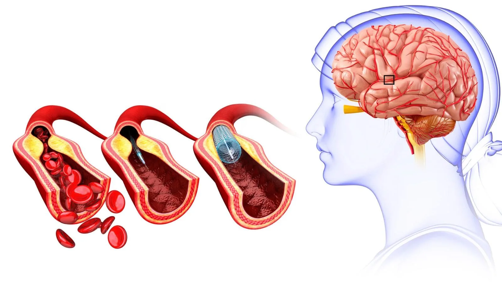
Sometimes the aneurysm neck is very wide. In such cases, there is a risk that the coils placed inside the sac may protrude back into the main vessel. This is where stent-assisted coiling comes into play. In this technique, before coiling, a flexible mesh stent is placed inside the main vessel across the aneurysm neck. This stent acts as a scaffold or cage, ensuring that the coils remain safely within the sac and preventing them from protruding outward. After the stent is placed, the microcatheter is passed through the stent struts and the aneurysm sac is safely filled. One stent may be used, or two stents may be used to protect two vessels (if their configurations are likened to letters: Y stent, T stent, H stent, etc.).
Flow-diverter stents represent an entirely different philosophy in aneurysm treatment. This method was developed especially for neckless fusiform aneurysms or giant aneurysms where coiling is difficult. The goal here is not to occlude the aneurysm sac but to redirect blood flow. Flow diverters are special stents with a much denser mesh structure than conventional stents. When placed in the vessel segment containing the aneurysm, they allow most of the blood to flow straight through the stent without entering the aneurysm. Blood flow into the aneurysm sac is dramatically reduced. Deprived of flow, the aneurysm gradually thromboses and shrinks over time. More importantly, the body eventually covers the stent with its own healthy vessel cells, effectively creating a new, smooth vessel wall in that area. This is not just an occlusion but a true biological healing and reconstruction.
Intrasaccular flow disruptors are the newest generation devices. Among the first to be implemented is the WEB device, a cage-like structure placed inside the aneurysm sac for not very large saccular aneurysms. Devices produced by different companies under names such as LUNA, Contour, SEAL, Trenza, and Medina, with varying shapes, are increasingly being used worldwide.
Should Open Surgery or Endovascular Methods Be Preferred in Aneurysm Treatment?
“Which treatment is better for me?” is the most fundamental question in the minds of every patient diagnosed with an aneurysm and their relatives. There is no single correct answer; the decision is made on an individual basis depending on the patient’s age, general health, and the aneurysm’s location, shape, and size. Both methods have their own advantages and disadvantages:
The main points considered when comparing the two approaches are:
- Recovery Process and Comfort
- Durability of Treatment and Recurrence Rate
- Patient Suitability
- Short-Term Risks
Open surgery (clipping) is a major operation in which the skull is opened. The recovery process is longer and more demanding, and hospital stays are longer. However, its greatest advantage is that it usually offers a definitive and permanent solution in a single procedure. A properly clipped aneurysm has a very low risk of recurrence years later.
Endovascular methods (coiling, stents, flow diverters), being closed procedures, are much more comfortable. There is no large surgical scar, hospital stays are much shorter (1–2 days for unruptured aneurysms), and patients return to daily life much faster. For elderly patients, those with additional health problems, or those whose general condition cannot tolerate major surgery, endovascular treatments are a much safer alternative. Large studies in ruptured aneurysms have shown that endovascular treatment is superior to open surgery in terms of short-term survival and reduced disability. However, the long-term recurrence risk of endovascular treatment, especially coiling, may be somewhat higher than that of surgery. Sometimes the coil mass may compact over time, allowing part of the aneurysm to refill with blood. Therefore, patients who undergo endovascular treatment may require periodic angiographic follow-up.
What Is the Difference Between Coiling and Flow Diverters in Endovascular Treatment?
There are also different philosophies within endovascular treatments themselves, which become particularly evident when dealing with complex aneurysms. Coiling and flow-diverter stents may enter through the same door, but they represent two distinct technologies aimed at entirely different goals. This difference can be likened to the contrast between “plugging a balloon” and “repairing a burst pipe.”
The main distinctions between the two methods are:
- Treatment Principle
- Suitable Aneurysm Type
- Healing Mechanism
- Long-Term Success
The basic principle of coiling is to mechanically fill the aneurysm sac and prevent blood entry. The target is directly the aneurysm balloon itself. This method works excellently for narrow-necked, saccular aneurysms. At the time of treatment, angiography shows immediate closure of the aneurysm.
The philosophy of the flow-diverter stent is entirely different. The target is not the aneurysm sac but the main vessel from which the aneurysm arises. The goal is to redirect blood flow away from the aneurysm, “starving” it, and to repair the diseased vessel wall using the body’s own healing mechanisms. For this reason, it is used especially in neckless, fusiform aneurysms or giant aneurysms where “the pipe itself” is problematic. The aneurysm does not close immediately at the time of treatment; blood flow slows, and complete closure may take weeks or even months. This is a biological healing process rather than a mechanical occlusion.
In the long term, flow diverters offer higher complete occlusion rates and much lower recurrence rates than coiling in the complex aneurysm group they treat. This is because they repair not just the symptom but the source of the problem—the diseased vessel wall. For this reason, flow-diverter technology is considered a groundbreaking approach that defines the future direction of endovascular treatment and represents a major milestone in aneurysm therapy.
Frequently Asked Questions
What Is an Aneurysm and how does it develop within blood vessels?
An aneurysm is an abnormal bulging or weakening of a blood vessel wall. It develops when the vessel loses structural strength, allowing it to expand under pressure and potentially increase the risk of rupture.
What Are the Symptoms of a Brain Aneurysm before it ruptures?
Many brain aneurysms cause no symptoms until they become large or rupture. Some individuals may experience headaches, vision problems, facial pain, or nerve-related symptoms due to pressure on nearby structures.
What Are the Symptoms of a Brain Aneurysm after rupture occurs?
A ruptured brain aneurysm often causes a sudden and extremely severe headache. Additional symptoms may include nausea, vomiting, neck stiffness, confusion, loss of consciousness, and neurological deficits.
Which risk factors increase the likelihood of developing a brain aneurysm?
Risk factors include high blood pressure, smoking, family history, advancing age, certain genetic disorders, excessive alcohol consumption, and conditions that weaken blood vessel walls over time.
Can a Brain Aneurysm exist without causing noticeable symptoms?
Yes, many brain aneurysms remain asymptomatic for years and are discovered incidentally during imaging studies performed for unrelated medical conditions. Regular evaluation may be recommended in selected cases.
How is a Brain Aneurysm diagnosed by healthcare specialists?
Diagnosis typically involves imaging studies such as CT scans, MRI, CT angiography, MR angiography, or cerebral angiography. These tests help identify the aneurysm’s size, location, and rupture risk.
What treatment options are available for a Brain Aneurysm?
Treatment depends on the aneurysm’s size, location, symptoms, and rupture risk. Options may include monitoring, endovascular coiling, flow diversion, surgical clipping, or other specialized interventions.
Can minimally invasive procedures be used to treat Brain Aneurysms?
Yes, many aneurysms can be treated through endovascular techniques such as coiling or flow-diverting stents. These minimally invasive procedures are performed through blood vessels without open brain surgery.
How long does recovery take after Brain Aneurysm treatment?
Recovery varies depending on whether the aneurysm ruptured and the treatment performed. Some patients recover within weeks, while others may require months of rehabilitation and neurological follow-up care.
Can a Brain Aneurysm be prevented through lifestyle changes?
Although not all aneurysms can be prevented, controlling blood pressure, avoiding smoking, maintaining a healthy lifestyle, and managing cardiovascular risk factors may help reduce the likelihood of aneurysm growth or rupture.

Prof. Dr. Özgür Kılıçkesmez is one of Türkiye’s leading interventional radiology and neuroradiology specialists, focusing on minimally invasive endovascular treatments. His primary areas of expertise include aneurysm and stroke treatments, embolization procedures, non-surgical treatments of thyroid and parathyroid diseases, interventional oncology, and peripheral vascular interventions. He currently serves as the Head of the Interventional Radiology and Neuroradiology Department at Istanbul Memorial Göztepe Hospital.
Graduating from Istanbul University Cerrahpaşa Faculty of Medicine in 1997, Prof. Dr. Kılıçkesmez received advanced training in interventional radiology and oncology as a Clinical Fellow at London Guy’s & St Thomas’ and King’s College Hospital between 2012 and 2013. He earned the title of Professor in 2020 and, in the same year, became the Founding Interventional Radiologist at SBÜ Çam and Sakura City Hospital. He also holds the EBIR (European Board of Interventional Radiology) certification.
Prof. Dr. Kılıçkesmez has 94 international and 40 national publications and over 2500 citations. He serves as a reviewer for prestigious journals such as the American Journal of Roentgenology (AJR) and Diagnostic & Interventional Radiology. With his clinical and academic expertise, he is a nationally and internationally recognized authority in stroke treatment, brain aneurysm, embolization, tumor ablation, and minimally invasive treatments of thyroid/parathyroid diseases.





Vaka Örnekleri