


Interventional oncology is a medical subspecialty that uses image-guided, minimally invasive procedures to diagnose and treat cancer. It focuses on targeting tumors directly with precision techniques such as ablation, embolization, and biopsy while preserving surrounding healthy tissues.
Image-guided cancer treatments such as tumor ablation and transarterial chemoembolization provide localized therapeutic effects by using radiologic imaging modalities. These procedures reduce systemic side effects and are commonly applied in liver, kidney, and lung malignancies.
Minimally invasive oncology procedures improve patient recovery time and reduce hospitalization compared to conventional surgery. Techniques performed under imaging guidance enable precise tumor targeting, resulting in lower complication rates and improved quality of life.
Role of interventional oncology in multidisciplinary cancer care involves collaboration with oncology, surgery, and radiology teams. It supports personalized treatment planning, especially in patients who are not suitable for surgery, offering effective palliative and curative options.
What Is Interventional Oncology and What Position Does It Hold in Medicine?
If we liken cancer treatment to a table, the legs of this table have for many years been surgery, chemotherapy, and radiotherapy. However, in recent years, a fourth and very powerful leg has been added to this table: Interventional Oncology. This discipline is the art of performing treatment by using the diagnostic power of radiology. We use imaging modalities such as ultrasound, tomography, or angiography devices like navigation systems (GPS).
These technologies show us the inside of the body, the vessels, the organs, and most importantly, the exact location of the tumor with millimetric precision. Guided by these images, we reach the tumor directly without cutting the skin, entering only through the blood vessels or through the skin with a needle. This is a “pinpoint” treatment. While in traditional methods the entire body may sometimes be exposed to treatment, in our methods the target is only the cancerous tissue. Interventional oncology is sometimes a life-saving alternative when surgery is not possible, and sometimes a powerful companion that enhances the effectiveness of other treatments.
What Does the Minimally Invasive Approach Provide for Interventional Oncology Patients?
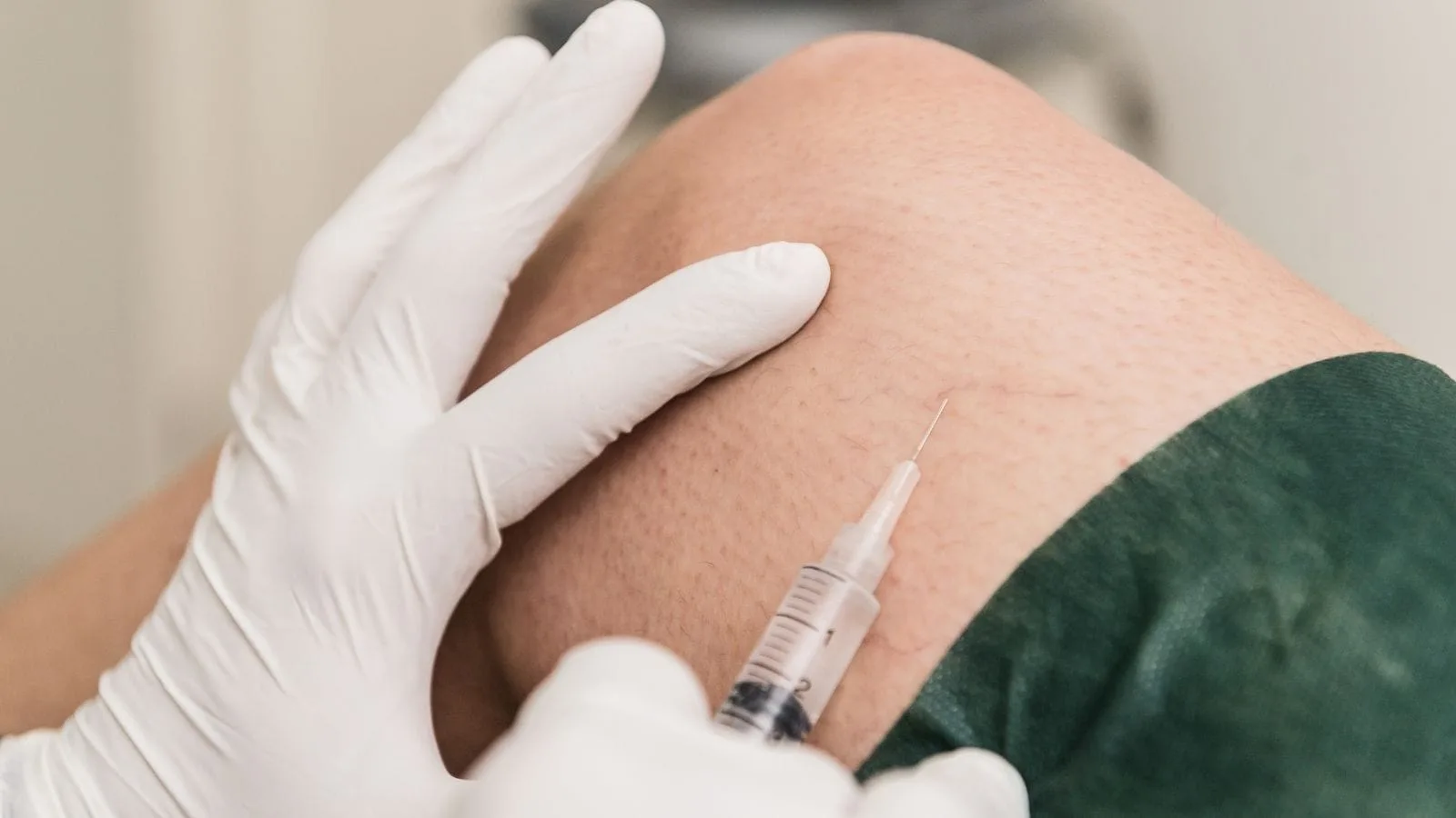
You often hear the term “minimally invasive” in medicine. Its meaning is “intervention that causes the least harm.” When treating a patient, we aim to minimize the physical damage and trauma we inflict. In open surgeries, large incisions are required for the surgeon’s hands to work. These incisions prolong the healing process, cause pain, and carry a risk of infection. In contrast, in our procedures the entry point is only the size of a needle hole. This entry point, usually 1–2 millimeters, does not even require sutures after the procedure; only a small bandage is applied.
The tangible benefits this approach provides to our patients are as follows:
- A strong supportive treatment for local masses that have not responded to medical oncological therapies
- Very little blood loss
- Minimal pain sensation
- Low risk of infection
- Possibility of rapid discharge
- Short recovery time
- No scarring from an aesthetic standpoint
- Early return to daily life
Especially for elderly patients or those with additional diseases such as heart or lung failure, for whom receiving general anesthesia is risky, these methods are literally a lifeline. Instead of staying in the hospital for days or weeks, being able to return home usually on the evening of the procedure or the next day has an incredibly positive effect on our patients’ morale and motivation.
Why Is Imaging Technology Vital in Interventional Oncology Procedures?
The heart of our work is imaging devices, which are our eyes and ears. A surgeon sees tissue with their eyes and feels it with their hands during surgery. We, on the other hand, see the tissues inside the body on our screens. That is why there is no such thing as a “blind” procedure for us. Devices such as Ultrasonography (US), Computed Tomography (CT), and Magnetic Resonance Imaging (MRI) provide us with a roadmap.
Thanks to the guidance provided by these devices, we can pass a very thin needle or catheter through the most complex pathways of the body and reach the target directly without damaging critical organs, major vessels, or nerves. For example, reaching a tumor located in the very center of the liver without touching the bile duct passing right next to it requires great skill and technology. Imaging guidance maximizes procedural safety. We see the answer to the question “Are we in the right place?” instantly on the screen. This increases the effectiveness of the treatment while reducing the risk of side effects to almost zero.
How Is the Biopsy Procedure for Cancer Diagnosis Performed by Interventional Oncology?
The first rule of fighting cancer is to know the enemy. When a mass is detected, starting treatment without knowing whether it is benign or malignant, and if malignant, what type it is and what its genetic characteristics are, is like shooting randomly in the dark. Therefore, to make a definitive diagnosis, it is essential to take a sample from that tissue, namely to perform a biopsy. In the past, patients sometimes had to undergo surgery even for a biopsy. Today, however, this procedure has become extremely practical in interventional radiology units.
The general steps we follow in the biopsy procedure are as follows:
- Application of local anesthesia for patient comfort
- Determination of the tumor location with an imaging device
- Entering through the skin with a special needle
- Taking millimetric tissue samples from inside the tumor
- Removing the needle and ending the procedure
We usually use special gun-type needles called “Tru-cut.” These needles allow us to obtain thin, thread-like tissue samples from the tumor. These samples are so valuable that they not only establish the name of the cancer, but also enable genetic tests that determine whether targeted therapies, which we call “Smart Drugs,” will work for that patient. In other words, that tiny piece draws the entire treatment roadmap for the patient.
What Is Tumor Ablation Treatment and How Does Interventional Oncology Perform It?
After the diagnosis is made, it is time for treatment. If the tumor is of suitable size and location, it may not be necessary to cut it out of the body. With the method we call “ablation,” we can destroy the tumor in its place. When explaining this to my patients, I sometimes use the analogy of “cooking an egg.” Just as an egg changes its structure and solidifies when heated, tumor cells also lose their vitality and die when exposed to a certain level of heat.
In this procedure, under imaging guidance, we place a special needle through the skin into the very center of the tumor. Then we activate the device connected to this needle. Using Radiofrequency (RFA) or Microwave (MWA) energy, we generate high heat at the tip of the needle. This heat destroys the tumor and the surrounding safety margin by burning them. When the procedure is finished, we withdraw the needle. Over time, the body clears that dead tissue, or it remains as harmless scar tissue. There is also the opposite method called “Cryoablation,” which is based on killing the tumor by freezing it. The tumor is destroyed by turning it into an ice ball.
In Which Organs Is the Ablation Method Successfully Used?
Ablation may not be suitable for every cancer type or every stage. However, in appropriate patients, it creates miraculous results. The most frequent and most successful organ of application is unquestionably the liver. Ablation is very effective in primary liver cancers or in metastases that have spread to the liver from organs such as the colon or breast.
Other organs in which ablation is successfully applied are as follows:
- Lung
- Kidney
- Bone
- Thyroid
- Adrenal gland
- Soft tissues
Especially in lung cancer patients who cannot tolerate surgery due to low lung reserve and limited breathing capacity, ablation can be life-saving. Similarly, in kidney tumors, it is possible to preserve the organ by burning only the tumor without removing the entire kidney. In bone metastases, the goal is sometimes not to destroy the tumor but to relieve the unbearable bone pain the patient is suffering from; interrupting nerve endings to stop the pain provides great comfort for the patient.
How Is a Tumor Starved by Interventional Oncology with Intravascular Treatments (TACE)?
In some cases, tumors are numerous or too large to be treated with ablation. At that point, we change strategy. Instead of hunting the enemy one by one, we decide to cut off its supply routes. Tumors are structures that grow very rapidly and constantly need energy. Therefore, they form new vascular networks to nourish themselves. We use the angiography method, enter through the groin vessel, and advance through the vascular map of the body. Our goal is to find the tiny artery that directly feeds the tumor.
In the “Chemoembolization” (TACE) procedure, we advance our catheter tip to the very doorstep of the tumor. From there, we deliver two things: first, a concentrated chemotherapy drug; second, micro-particles that block the vessel. This procedure creates a dual effect.
The effects of TACE on the tumor are as follows:
- Poisoning with a high-dose drug
- Depriving it of oxygen
- Cutting off its nutrient supply
- Stopping its growth
- Causing it to shrink
Normally, chemotherapy given through the arm spreads throughout the body and causes side effects such as hair loss and severe nausea. In the TACE procedure, since we trap the drug directly in the tumor and also block the vessel, the drug mixes very little with the systemic circulation. Thus, while the patient experiences far fewer of the severe side effects of chemotherapy, the tumor is exposed to a much higher dose of the drug. This method is the standard treatment especially for inoperable liver cancers.
How Does Interventional Oncology Use Radiation with Radioembolization (Y-90) Therapy?
Radioembolization (TARE), sometimes popularly known as “atomic therapy” or “pinpoint irradiation,” is one of the latest marvels of technology. In this method, we again enter through the groin using angiography. However, this time we do not send chemotherapy drugs into the tumor vessel, but millions of microscopic spheres loaded with a radioactive substance (Yttrium-90).
These spheres are so small that they advance into even the finest capillaries within the tumor and lodge there. From where they settle, they emit very powerful radiation over an area of only a few millimeters. In external radiotherapy, radiation beams may affect healthy tissues as they pass through the skin before reaching the tumor. In this method, however, we place the radiation source directly “inside” the tumor itself. In this way, while healthy liver tissue is preserved, the tumor is destroyed by being irradiated from within.
The situations in which radioembolization is most frequently preferred are as follows:
- Tumors resistant to chemotherapy
- Very large masses
- Patients with portal vein thrombosis
- Cases not suitable for surgery
- Situations where the TACE procedure is risky
This method is a kind of “Trojan Horse” tactic. We infiltrate the enemy fortress (the tumor) and start the battle from the inside. Especially in difficult patients in whom TACE cannot be performed due to obstruction of the main liver vessels, this method is our most powerful and sometimes the only option.
Which Supportive Procedures Are Performed to Improve the Quality of Life of Cancer Patients?
Interventional Oncology does not focus solely on destroying the tumor. For us, the patient is not just a tumor; they are a human being with pain, comfort needs, and quality of life. Cancer treatment is a long and challenging marathon. Making the patient’s life easier during this process is also our duty. For example, in patients who will receive continuous chemotherapy, arm veins may thin out and rupture over time, turning venous access into torture.
To prevent this, we perform the “Port Catheter” procedure. We place a small chamber (port) under the skin, usually in the chest area. This chamber is connected to a central vein. The patient can receive all treatments and have blood drawn through this port without any pain.
The advantages provided by a port catheter for patients are as follows:
- Elimination of the struggle to find a vein
- No bruising on the arms
- Safe administration of medications
- Reduced risk of infection
- No interference with bathing
- Usable for many years
In addition, in advanced-stage cancers, tumors may sometimes compress the bile ducts or urinary tracts. In this case, the patient becomes jaundiced, experiences itching, or the kidneys approach failure. We use non-surgical methods again, entering only through needle-sized openings, to place a “catheter” or “stent” into these blocked pathways. By opening the obstruction, we restore bile or urine flow. Even if these procedures do not eliminate the cancer, they save the patient’s organs, prevent intoxication, and allow them to spend their remaining life with much higher quality. Pain management is equally important; relieving severe cancer-related pain by performing nerve blocks and easing the patient’s suffering is one of the most humane aspects of the art of medicine.
How and by Whom Are Treatment Decisions Made in Interventional Oncology?
Cancer treatment is a process too complex for a single doctor to decide alone. The mentality of “I did it, it’s done” has no place in modern medicine. Each patient’s disease is unique like a fingerprint and requires individualized planning. Therefore, there is a structure called the “Multidisciplinary Tumor Board.” In this board, the medical oncologist (drug therapy specialist), radiation oncologist (radiation therapy specialist), surgeon, pathologist, and we interventional radiologists gather around a table.
We collectively evaluate the patient’s imaging studies, laboratory tests, biopsy results, age, comorbidities, and even social situation. We seek an answer, through collective reasoning, to the question: “Is the best option for this patient surgery, medication, or ablation?” As interventional radiologists, at this table we present minimally invasive options for patients who are not suitable for surgery or who will not benefit from surgery.
The goals of the board are to ensure:
- Selecting the most appropriate treatment
- Preventing loss of time
- Avoiding unnecessary procedures
- Approaching the patient holistically
- Prolonging survival
- Preserving quality of life
Sometimes we start treatment and shrink the tumor, and then the surgeon operates. Sometimes surgery is not possible, and we step in to control the disease. This team effort is of vital importance for the patient’s success.
Frequently Asked Questions
What is interventional oncology and how does it differ from traditional cancer treatment?
What types of cancers can be treated with interventional oncology procedures?
How do image-guided treatments work in interventional oncology?
What are the most common procedures performed in interventional oncology?
Who is a suitable candidate for interventional oncology treatments?
What are the advantages of interventional oncology compared with open surgery?
Can interventional oncology be combined with chemotherapy or radiation therapy?
What risks and complications are associated with interventional oncology procedures?
How long does recovery take after an interventional oncology procedure?
What experiences do patients commonly report after interventional oncology treatment?

Prof. Dr. Özgür Kılıçkesmez is one of Türkiye’s leading interventional radiology and neuroradiology specialists, focusing on minimally invasive endovascular treatments. His primary areas of expertise include aneurysm and stroke treatments, embolization procedures, non-surgical treatments of thyroid and parathyroid diseases, interventional oncology, and peripheral vascular interventions. He currently serves as the Head of the Interventional Radiology and Neuroradiology Department at Istanbul Memorial Göztepe Hospital.
Graduating from Istanbul University Cerrahpaşa Faculty of Medicine in 1997, Prof. Dr. Kılıçkesmez received advanced training in interventional radiology and oncology as a Clinical Fellow at London Guy’s & St Thomas’ and King’s College Hospital between 2012 and 2013. He earned the title of Professor in 2020 and, in the same year, became the Founding Interventional Radiologist at SBÜ Çam and Sakura City Hospital. He also holds the EBIR (European Board of Interventional Radiology) certification.
Prof. Dr. Kılıçkesmez has 94 international and 40 national publications and over 2500 citations. He serves as a reviewer for prestigious journals such as the American Journal of Roentgenology (AJR) and Diagnostic & Interventional Radiology. With his clinical and academic expertise, he is a nationally and internationally recognized authority in stroke treatment, brain aneurysm, embolization, tumor ablation, and minimally invasive treatments of thyroid/parathyroid diseases.




Vaka Örnekleri