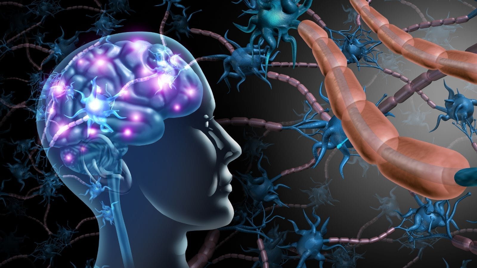
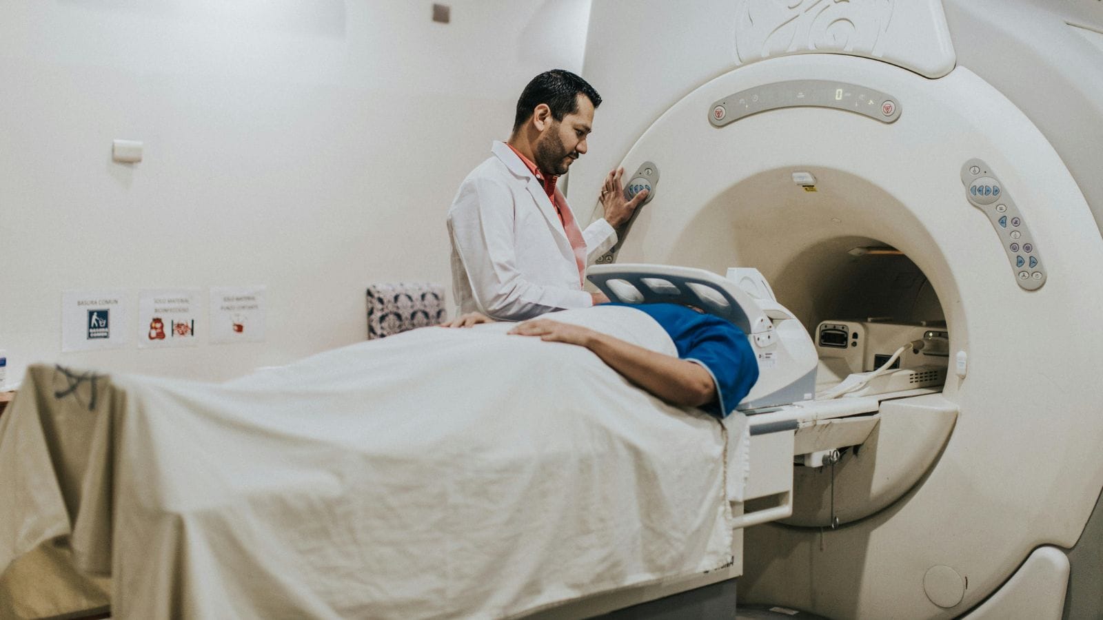
Malformation refers to structural abnormalities in organs or tissues that develop during the embryonic stage. These defects may affect the shape, size, or function of an organ, and can range from mild to severe, depending on the affected system.
Causes of malformation include genetic mutations, chromosomal disorders, and environmental factors. Exposure to infections, toxins, or nutritional deficiencies during pregnancy may also contribute to developmental abnormalities.
Common malformations include cleft lip, congenital heart disease, and neural tube defects. Early detection through prenatal imaging and genetic testing allows timely intervention and counseling for families.
Treatment depends on severity and location. Some malformations require surgical correction after birth, while others may be managed with medical therapy, rehabilitation, or long-term supportive care.
What Is Malformation and How Is It Medically Defined?
Malformations are usually examined under the heading of “congenital anomalies” in medical literature. The word “congenital” means present at birth or during delivery. Thus, a malformation refers to a structural or sometimes functional abnormality that is already present when the baby is born. However, malformations do not necessarily have to be apparent at birth; anomalies that begin during fetal development and become more prominent later are also included in this category.
These structural abnormalities can occur anywhere in the body: Improper folding of the brain (e.g., lissencephaly), improper separation of heart chambers (e.g., ventricular septal defect), or open spine (e.g., spina bifida) are examples that show how broad this concept is. The key point is that the malformation directly affects a “structural formation” in the body. Functional disorders can also occur, but at its core, there is a “morphological” issue.
To use a simple analogy, think of a puzzle prepared for a child. If the pieces are not placed correctly, part of the picture will look missing or flawed. Similarly, in malformations, problems occur during the placement, shaping, or differentiation of “pieces,” i.e., cells and tissues. If a piece is misplaced or missing, a visible defect appears in the whole.
Medically, malformations are classified as “minor” and “major.” Minor malformations (such as a small indentation in the ear, an extra crease in the hand, or a small skin fold) usually do not pose a life risk or cause significant loss of function. However, “major” malformations (such as severe heart anomalies, neural tube defects, or major skeletal abnormalities) can deeply affect daily life or be life-threatening.
What Are the Main Types of Congenital Malformations?
Congenital malformations are essentially divided into structural and functional types, but the subcategories are quite diverse and complex.
Cardiovascular Malformations
- This group is among the most common of all congenital anomalies. Holes between the heart chambers (e.g., atrioventricular septal defect), incorrect positioning of vessels (e.g., transposition of the great arteries), or failure of vessels that should close after birth (e.g., patent ductus arteriosus) fall into this class.
- In some babies, symptoms like heart murmur, cyanosis, or shortness of breath may occur. Treatment generally involves surgical procedures or catheter-based corrections.
Nervous System and Neural Tube Defects
- Neural tube defects involving the development of the brain and spinal cord are included here. Spina bifida, anencephaly, or underdeveloped brain folds (lissencephaly) are typical examples.
- Such anomalies can often be detected in early pregnancy ultrasounds. Some carry life risks, while others may be partially corrected with surgery.
Craniofacial Malformations
- The most well-known example is cleft lip and palate. If the facial structures do not join properly between weeks 5 and 12 of pregnancy, this occurs.
- Cleft lip and palate can cause difficulties in feeding and speech, but surgical techniques now offer a high chance of correction.
Musculoskeletal System Malformations
- This wide group can affect limbs or the spine. Joint disorders (such as hip dysplasia), extra fingers (polydactyly), or limb reduction defects are included.
- Some can be diagnosed very early, even in the womb. Surgical treatment, physical therapy, or special orthotic-prosthetic methods may be used.
Urogenital Malformations
- This includes conditions such as renal agenesis (absence of kidneys) or abnormal opening of the urinary tract (e.g., hypospadias).
- Symptoms after birth may include kidney function problems, frequent urinary tract infections, or urination difficulties due to anatomical defects. Surgical repair or long-term medical treatment may be needed.
Functional/Metabolic Anomalies
- Here, instead of a structural abnormality, there is a disruption in chemical processes or hormones. Disorders like phenylketonuria (a protein metabolism defect) or congenital hypothyroidism may not be visible externally but cause significant dysfunctions.
- With early diagnosis (especially newborn screenings), healthy lives can be achieved with medication or special diets.
These types sometimes occur alone, or multiple malformations may be present in the same baby. For example, in chromosomal disorders like Down syndrome (trisomy 21), several anomalies involving the heart, digestive, and skeletal systems may be found together.
How Do Genetic Factors Contribute to Malformations?
Our genetic inheritance is like an architect’s project drawings. A small error in the plan can cause mistakes in the “construction” of the body. Genetic factors can be examined in three main groups:
Chromosomal Anomalies
- Chromosomes are DNA packages in the cell nucleus. Abnormalities in their number or structure can lead to various malformations. For example, an extra chromosome (trisomy) or a missing one (monosomy) can seriously affect development.
- Down syndrome is a well-known example, characterized by an extra 21st chromosome (three copies in total), with symptoms like heart anomalies, characteristic facial features, growth, and intellectual disabilities.
Single-Gene Disorders (Mendelian Inheritance)
- Sometimes the issue is not in the entire chromosome but in a specific gene mutation. In disorders such as phenylketonuria, cystic fibrosis, or sickle cell anemia, the faulty gene directly disrupts a body function, leading to malformation or functional abnormality.
- Such errors can be inherited or appear for the first time as new (de novo) mutations in an individual.
Changes in Regulatory DNA Regions
- Some genes act as “managers” controlling where, when, and how much other genes work. Mutations in these regulatory regions can result in abnormalities such as improper limb growth or abnormal blood vessel formation.
- In some arteriovenous malformations, these regulatory factors are thought to be involved, although many of these mechanisms are not yet fully understood.
Which Environmental Factors Cause Malformations?
The effect of environmental factors can be imagined as foreign substances falling onto fresh dough. If something contaminates the dough before baking, shape defects or unwanted tastes may emerge during baking. For the fetus, harmful external factors can disrupt the development process.
Chemicals and Toxic Substances (Teratogens)
- Elements like lead, mercury, arsenic, pesticides, industrial solvents, or radiation can severely disrupt cell division and tissue formation in the fetus.
- For example, exposure to high doses of radiation during pregnancy can cause problems in the baby’s brain development and bone marrow formation.
Drugs and Other Pharmaceutical Agents
- Some drugs are strictly contraindicated in pregnancy because they have teratogenic effects (e.g., isotretinoin).
- Certain anticonvulsants (such as valproic acid) used for epilepsy are known to increase the risk of neural tube defects. Women planning pregnancy should have their medication regimen reviewed by a doctor.
Infections (TORCH Infections)
- Toxoplasma, rubella, cytomegalovirus (CMV), herpes simplex virus (HSV), and similar infections can cause serious organ damage in the baby, especially if contracted in the first trimester. For example, rubella exposure can result in heart anomalies, cataracts, and hearing loss.
- Zika virus infection can cause severe brain malformations such as microcephaly (small head and brain).
Lifestyle and Nutrition Habits
- Smoking, alcohol consumption, or poor nutrition reduce the amount of oxygen and nutrients delivered to the baby through the placenta, leading to deficiencies in the “building materials” required for organ development. Alcohol can cause “Fetal Alcohol Syndrome” with facial anomalies and intellectual disabilities.
- Folic acid deficiency is a leading factor increasing the risk of neural tube defects, so folic acid supplementation is recommended from the planning stage of pregnancy.
Physical Factors and Work Environment
- Standing for long periods, heavy lifting, working in high heat, or close to radiation sources can also pose risks during pregnancy. Although mechanisms are not fully explained, studies suggest an increase in neural tube defects among those working in radioactive environments.
Can Malformations Be Detected During Pregnancy?
Today’s advances in medical technology allow many malformations to be detected before birth. Early diagnosis enables necessary precautions. Two main methods are prominent:
Prenatal Ultrasonography
- Routine ultrasound scans at different stages of pregnancy assess the growth and development of the baby’s organs. The first trimester (11–14 weeks) scan can detect evident brain defects or severe heart problems, but a more detailed examination is usually done between weeks 18–22.
- The second trimester scan can provide a clearer view of the baby’s heart chambers, spine, kidneys, gastrointestinal system, and facial structure. For example, cleft lip/palate, abdominal wall defects (omphalocele, gastroschisis), or serious skeletal issues can be identified at this stage.
Additional Imaging and Tests
- Sometimes, ultrasound does not provide enough information, or a more detailed examination is required. In these cases, fetal echocardiography (for heart anomalies), MRI (for brain and spine details), amniocentesis, or chorionic villus sampling (for chromosomal analysis) may be performed.
- Blood tests also offer clues. For example, high levels of alpha-fetoprotein (AFP) in the blood may indicate neural tube defects. If a suspicious situation is detected, further imaging is used to confirm.
- Of course, not all malformations can be detected during pregnancy. The baby’s position, the limitations of ultrasound technology, and the mother’s body structure can affect screening success. However, in general, especially major and life-threatening anomalies can be detected before birth, providing a valuable advantage for postnatal intervention planning and informing parents.
What Are Arteriovenous Malformations (AVM) and How Do They Occur?
Arteriovenous malformations (AVMs) are abnormal “short circuits” that occur when the thin capillary network, which should normally be present between arteries and veins, is absent or underdeveloped. You can imagine this as a main water pipe connecting uncontrollably to the main reservoir of a city without the small pipes (capillaries) to regulate pressure. Without these small pipes, there is excessive pressure difference between the main pipe and the reservoir.
- Development Process: If, during the early stages of pregnancy when the vascular network forms, arteries and veins do not “bridge” correctly, these abnormal connections develop.
- Common in Brain and Spinal Cord: AVMs are particularly found in the brain and spinal cord. These vascular tangles can enlarge over time, press on brain tissue, or create a risk of hemorrhage. When bleeding occurs, severe clinical situations like stroke or seizures can arise.
- Delayed Symptoms: Many people are unaware they have an AVM for years, as small AVMs may not cause symptoms. However, as they grow, headaches, seizures, visual disturbances, or sudden and life-threatening brain hemorrhage can occur.
- Causes: Largely attributed to random somatic mutations or embryonic vascular formation errors, but may also be associated with some hereditary syndromes (e.g., Hereditary Hemorrhagic Telangiectasia).
What Are the Symptoms of Malformations in Newborns?
In newborn babies, some malformations are easily visible (such as cleft lip and palate, clear limb deficiencies), while others involving internal organs may be missed. Symptoms vary according to the affected organ:
Heart Anomalies
- Cyanosis (especially on the lips and nail beds)
- Fatigue or shortness of breath during feeding
- Heart murmur
- Inadequate weight gain, growth retardation
Nervous System Malformations
- Prominent or abnormal fontanelle
- Excessive muscle stiffness or floppiness (hypotonia or hypertonia)
- Lack of movement, poor response to stimuli
- Seizures
Craniofacial Malformations
- Facial asymmetry, ear shape abnormalities
- Feeding difficulty or milk coming from the nose due to cleft lip and palate
- Marked anomalies in the eyes or nose
Digestive System Malformations
- No meconium passage in the first 24–48 hours after birth (sign of bowel obstruction)
- Vomiting, bile-stained vomiting (green color)
- Abdominal swelling or abnormal bowel sounds
Urinary System Malformations
- Difficulty urinating or very infrequent urination
- Blood in urine or signs of infection
- Obvious swelling, such as a kidney mass on the abdomen
Limb and Skeletal System Disorders
- Extra fingers, obvious abnormalities of the hands or feet
- Limited joint movement, bone protrusions
- Postural abnormalities due to neck or spinal problems
How Are Malformations Diagnosed and Treated?
The diagnosis of malformations is made in both the prenatal and postnatal periods. While methods such as ultrasound, MRI, and genetic testing are used before birth, physical examination, imaging, and laboratory tests come into play after birth.
Diagnostic Methods
- Prenatal Ultrasound: Second-trimester detailed ultrasound is very effective for detecting major anomalies in the heart, abdominal wall, spine, and facial structure.
- MRI and Echocardiography: Used especially for brain and heart anomalies when ultrasound does not provide clear results. Fetal echocardiography is used to examine the structural details of the heart.
- Genetic Tests (Amniocentesis, Chorionic Villus Sampling): These are invasive methods used when chromosomal or single-gene disorders are suspected. The samples obtained are analyzed in detail in genetic laboratories.
Treatment Methods
- Surgical Intervention: Closing heart defects, repairing cleft palate, or spinal surgery are main approaches that can provide definitive solutions in some malformations. In some cases, surgery can be performed even while the baby is still in the womb (fetal surgery).
- Medical Treatments: In metabolic disorders (e.g., congenital hypothyroidism), replacing the missing hormone or enzyme is the main form of treatment. Supportive drug therapies are also used in some syndromic cases.
- Rehabilitation and Supportive Applications: For orthopedic malformations and neurological problems, physical therapy, occupational therapy, and special education are essential for long-term success.
- Endovascular and Radiosurgical Procedures: Especially for vascular malformations (such as AVMs), intravascular therapies or radiosurgery methods (such as Gamma Knife) may be preferred instead of classic surgery.
What Is the Difference Between Malformations and Deformations?
- Malformation: These are congenital abnormalities that arise from improper embryonic or fetal development. In other words, there is a problem in the “building blocks” during their construction.
- Deformation: This occurs when normally healthy tissue or organ takes on an abnormal shape due to mechanical, physical, or environmental influences in the later stages of pregnancy or after birth. For example, compression in the womb (especially in oligohydramnios) can cause shape abnormalities such as clubfoot.
Craniosynostosis caused by early fusion of skull bones (e.g., Apert syndrome) is a “malformation,” as the problem originates in the genetic or embryonic code that develops the skull bones. In contrast, if a baby develops flattening at the back of the head due to lying in the same position (positional plagiocephaly), this is a “deformation.” Here, the basic structure is normal, but external factors alter the form.
Deformations can usually be corrected or improved with simple physical measures (positioning, physical therapy) or orthoses. Malformations generally require more substantial surgical or medical interventions. This distinction is important for determining the correct treatment approaches.

Interventional Radiology and Neuroradiology Speaclist Prof. Dr. Özgür Kılıçkesmez graduated from Cerrahpaşa Medical Faculty in 1997. He completed his specialization at Istanbul Education and Research Hospital. He received training in interventional radiology and oncology in London. He founded the interventional radiology department at Istanbul Çam and Sakura City Hospital and became a professor in 2020. He holds many international awards and certificates, has over 150 scientific publications, and has been cited more than 1500 times. He is currently working at Medicana Ataköy Hospital.


Vaka Örnekleri