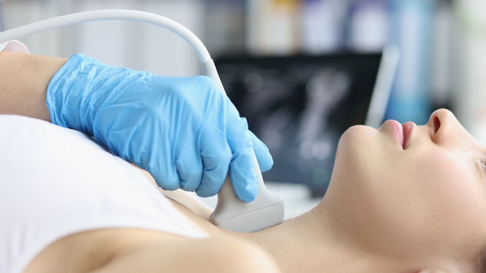
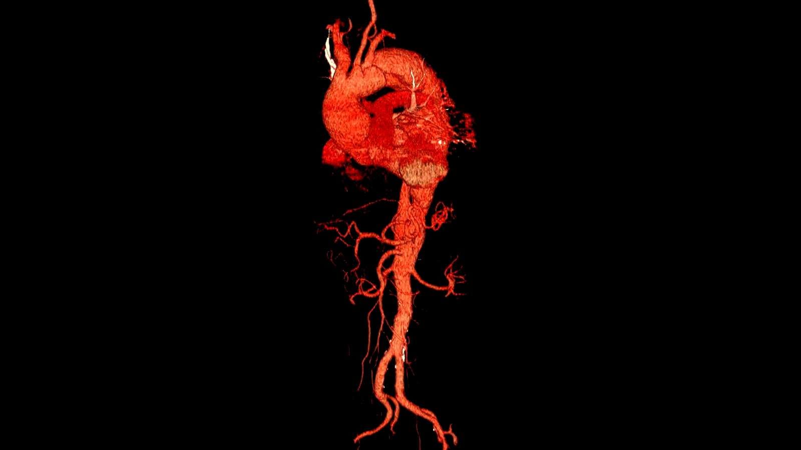
Radiology is a medical specialty that uses imaging technologies such as X-rays, CT, MRI, and ultrasound to diagnose and treat diseases. It plays a crucial role in modern medical care.
Diseases diagnosed with radiology include cancer, cardiovascular conditions, musculoskeletal disorders, neurological diseases, and infections. Imaging helps detect early changes not visible in physical exams.
Interventional radiology provides minimally invasive treatments such as embolization, biopsies, and ablations, reducing the need for open surgery. It combines imaging precision with therapeutic interventions.
Radiology ensures accurate diagnosis, treatment planning, and follow-up monitoring. It remains essential for effective patient care across nearly all medical specialties.
| Definition | Radiology is a medical science that uses methods such as X-rays, sound waves, and magnetic fields for the diagnosis and treatment of diseases. |
| Subfields | Diagnostic Radiology: Diagnoses diseases using imaging methods such as X-ray, CT, MRI, ultrasound. Interventional Radiology: Applies minimally invasive procedures (angiography, biopsy, tumor treatments, etc.) under imaging guidance for diagnosis and treatment. |
| Methods Used | X-ray: Used to evaluate bone fractures, lung diseases, and intestinal obstructions. Computed Tomography (CT): Used for detailed cross-sectional imaging (tumors, internal organ injuries, vascular diseases). Magnetic Resonance (MR): Uses a powerful magnetic field to image soft tissue, musculoskeletal system, brain, and spinal cord diseases. Ultrasonography (USG): Examines internal organs, pregnancy, and vascular structures using sound waves. Nuclear Medicine Imaging: Evaluates organ function using methods such as Positron Emission Tomography (PET) and scintigraphy. |
| Areas of Use | Detection of fractures and dislocations, cancer diagnosis, vascular diseases, internal organ diseases, pregnancy follow-up, brain and spinal cord diseases, interventional radiological treatments. |
| Risks and Side Effects | X-ray and CT: Radiation exposure may pose a long-term risk. MR: The magnetic field is risky for patients with metal implants. Contrast Agents: May cause side effects in patients with kidney failure. |
| Who Is Not Suitable? | Pregnant women (careful with radiation-containing methods), patients with kidney failure (for contrast agents), patients with pacemakers or metal implants (for MRI). |
What is Radiology and How Does It Use Imaging Technology to Diagnose Diseases?
Radiology is essentially the science of imaging internal structures of the body using various physical agents such as X-rays, magnetic fields, sound waves, or radioactive materials.
For example, X-ray is one of the oldest and most common methods. A low dose of X-ray passes through the body and is detected by a special sensor; dense tissues such as bone block the rays and appear white on film or digital screen. This makes it much easier to detect broken bones or see abnormalities in organs such as the lungs or intestines.
If X-ray is like taking a “black-and-white photo” of a specific area, Computed Tomography (CT) is like photographing a sculpture from different angles to create a three-dimensional model. CT scans combine X-ray slices taken from various angles and create cross-sectional images. This helps reveal even small tumors, cysts, infections, or bleeding in detail.
Magnetic Resonance Imaging (MRI) maps the body using magnetic fields and radio waves. It’s not as fast as X-ray or CT for hard tissues but is very successful at showing the structure of soft tissues and detailed features of vital organs such as the brain and spinal cord. Small tears in muscles and ligaments, brain tumors, or spinal compressions can be detected more easily with MRI.
Unlike the others, ultrasound uses high-frequency sound waves. Think of a street vendor announcing with a megaphone on a hot summer day; the sound spreads and reaches our ears. Ultrasound works on a similar principle, but with much higher frequency sound waves. The waves that reflect off tissues form an image on the screen. Because it does not involve radiation and provides real-time information, it is commonly used for pregnancy monitoring and heart and vascular examinations (such as using Doppler ultrasound to detect vascular occlusions).
Nuclear medicine methods (such as PET scans) involve administering radioactive tracers into the body. These tracers accumulate in the target organ or tissue and show the metabolic activity of that area. Cancer cells proliferate rapidly, so the tracer accumulates more in areas with cancer, revealing the location and size of the tumor. This technique is used to determine the stage and spread of cancer, assess heart muscle viability, or monitor brain activity in some neurological diseases.
Why Is Radiology Considered a Key Diagnostic Tool in Modern Medicine?
Today, early and accurate diagnosis is one of the main goals of medicine. Radiology is a key tool in achieving this goal. For example, for a patient presenting to the emergency room with signs of a heart attack, a doctor can quickly order a chest X-ray or CT angiography to check for blood clots in the pulmonary arteries or abnormal enlargement of the heart. Radiological imaging enables doctors to get fast answers to critical questions.
Modern medicine values details in understanding the nature of disease. Simply saying “the patient is in pain” is not enough. It’s crucial to know which tissue or organ is causing the pain. Radiological images provide clear and visual answers to these questions. Radiology is indispensable in cancer screening. For example, “mammography” can detect breast cancer at a very early stage, and low-dose CT is used to screen for lung cancer in high-risk groups. These techniques allow for the detection of critical diseases even before symptoms appear.
Radiology also helps physicians make fast and accurate decisions. A trauma patient with suspected bleeding can be examined by CT within minutes to determine if surgery is needed and which approach should be taken. This speed and accuracy show why radiology is so central in modern medicine.
How Does Radiology Help in Early Detection of Diseases?
The ideal way to treat a disease is to detect it as early as possible. Early diagnosis reduces mortality and makes treatment easier. Radiology’s role in this process is like detecting a small water leak in your home before it causes major damage. If caught early, it’s a small repair; if missed for months, it can damage the whole building.
For example, mammography can detect breast tumors just a few millimeters in size, enabling intervention before the mass grows or spreads. Low-dose CT scans can identify small nodules in high-risk patients for lung cancer. Early detection allows for more effective surgery or treatments.
Radiology is also crucial in the early diagnosis of some brain diseases. MRI can detect small brain tumors or vascular abnormalities before symptoms appear. Similarly, cysts and tumors in internal organs such as the liver, kidney, or pancreas can be found at an early stage. Like a car’s “check engine” light, radiology helps control disease before it gets worse, offering patients a chance for a longer, higher-quality life.
What Is the Role of Radiology in Monitoring Treatment and Evaluating Outcomes?
After diagnosis, the next step is to plan and closely monitor treatment. Radiology plays a key role here, guiding both doctor and patient. Just like using a navigation system to track your journey, radiological images allow observation of the disease’s course at every stage of treatment.
For example, consider a cancer patient undergoing chemotherapy or radiotherapy. CT or MRI scans are taken at intervals to assess if the tumor is shrinking or still growing. If the treatment is not working, different protocols can be evaluated. Similarly, for a patient with a vascular stent, ultrasound or angiography can check if the stent remains open. If there’s a narrowing, early intervention is possible.
Radiology can also detect possible complications in advance. After bone surgery, for example, regular X-rays monitor bone healing and check for problems such as non-union or malunion. Monitoring treatment effectiveness and safety is critical for long-term health, and radiology is the guide that provides clarity and reliable data for both physicians and patients.
How Do Different Imaging Techniques in Radiology Improve Disease Management?
- X-Ray: Fast, practical, and instantly accessible in most hospitals, it is the basic imaging tool for fractures, joint problems, lung diseases, or dental issues.
- Computed Tomography (CT): Produces detailed cross-sectional images, ideal for complex regions such as the abdomen, chest, and brain to quickly identify tumors, bleeding, or vascular problems.
- Magnetic Resonance Imaging (MRI): Acts like a detective for soft tissue and nervous system. Provides high-resolution images for brain, spine, muscles, ligaments, and cartilage.
- Ultrasound: No radiation, provides real-time imaging, and is cost-effective—preferred for pregnancy follow-up, abdominal organ evaluation, and cardiovascular assessments.
- Nuclear Medicine (PET, scintigraphy, etc.): Excels at creating a metabolic and functional map of the body, showing how tissues are functioning, especially in cancer, heart, and some neurological diseases.
Choosing the appropriate method ensures the most efficient outcome in diagnosis and treatment. Sometimes, multiple techniques are combined for a more complete assessment. New technologies, such as artificial intelligence-assisted image analysis, can now detect subtle details that might otherwise be missed, making radiology central not just to “seeing,” but to “understanding” as well.
Why Is Radiology Essential for Predicting Health Outcomes?
Health outcomes relate to the likely course of a disease and what results can be expected after treatment. Radiological images are among the strongest sources of data for making such predictions.
Just as a civil engineer inspects the structure of a building to assess its safety, radiology reports on the condition of tissues and organs. For example, a DEXA scan can measure bone density to predict the risk of fractures, or CT/MRI can track cancer recurrence or the change in lesions over time.
With developments in artificial intelligence and big data analysis, radiology now helps not only in visualizing but also in predicting outcomes. Radiomic analysis, for example, uses the features of tumors or lesions in images to estimate their future behavior, contributing to more personalized medicine.
What Are the Benefits of Radiology in Emergency and Primary Care?
Radiology is not just for major hospitals or severe cases. It is vital in emergencies and primary care (family medicine).
Emergencies: Trauma patients (traffic accidents, falls) can be quickly scanned for internal bleeding, brain hemorrhage, or organ rupture. A few minutes of CT scanning can provide doctors with life-saving information. Similarly, chest X-ray can instantly help diagnose pneumonia or other lung conditions. Imaging of the heart vessels (angiography or CT angio) enables rapid decision-making in suspected heart attacks.
Family medicine: Basic ultrasound or X-ray in the family doctor’s office can clarify many initial diagnoses. For example, ultrasound can reveal kidney stones in a patient with urinary complaints. Regular imaging in chronic diseases (such as chest X-ray for asthma or COPD) helps track changes and disease progression.
How Do Doctors Use Radiology to Understand the Causes of Symptoms?
When you hear a patient’s symptoms, many possible diagnoses come to mind. Radiology makes life easier for both doctors and patients by providing visual evidence. The choice of imaging method is based on the likely cause; for example, right lower abdominal pain may lead to ultrasound for suspected appendicitis, but if the image is unclear, CT may be used. Similarly, back pain may require X-ray or MRI to distinguish between spinal or kidney problems.
Every imaging method has its own advantages, and the physician’s knowledge of when to use which technique ensures targeted diagnosis rather than random searching. Once the underlying cause is clarified, the best treatment plan can be offered.
Why Is Precision Critical in Radiology for Accurate Diagnosis and Treatment?
“Accurate diagnosis brings accurate treatment” is a key principle in medicine. Radiology is crucial for revealing the real nature of disease. The accuracy of imaging and its interpretation is very important. For example, movement during an MRI or improper machine settings may result in blurred or misleading images, while incorrect targeting in X-ray may miss important details.
Precision matters not only in technical aspects but also in interpretation. Radiologists systematically review images to detect even small lesions. Artificial intelligence software can now assist in identifying subtle findings that may be missed by the human eye. For instance, a tiny nodule on a lung X-ray could signal early-stage lung cancer and be life-saving if detected.
Incorrect or incomplete interpretation may result in overtreatment or delay in necessary therapy. Missing a tumor could allow a disease to progress, while mistaking a benign lesion for a malignancy could lead to unnecessary interventions and anxiety. Therefore, precision in radiology is essential from start to finish for safe and reliable healthcare.
FAQ
- What are radiological examinations?
Radiological examinations include various methods used to image the internal structure of the body. The most common are X-ray, computed tomography (CT), magnetic resonance imaging (MRI), ultrasonography (USG), and nuclear medicine imaging (PET, scintigraphy). Each uses different technologies to provide specific information. - What are the types of radiology?
Radiology is mainly divided into three branches: Diagnostic Radiology (for interpreting images for diagnosis), Interventional Radiology (for non-surgical treatments and diagnostic procedures under imaging guidance), and Radiation Oncology (for treating cancer with radiotherapy). - Why do people go to the radiology department?
People visit radiology for many reasons, most commonly for imaging to diagnose a disease (e.g., suspected fracture or internal organ disease). Radiology is also used to manage treatment (such as interventional radiology), to screen for diseases like cancer, and to monitor post-treatment status. - What is the difference between medical imaging and radiology?
Medical imaging is the general process of making the internal structure of the body visible through methods such as X-ray, MRI, ultrasound, etc. Radiology is the medical specialty that uses these imaging methods to diagnose and sometimes treat diseases. In other words, medical imaging is the core tool of radiology. - What devices are used in radiology?
Different devices are used depending on the area and purpose: X-ray machines, CT scanners (for cross-sectional imaging with X-rays), MRI machines (with strong magnets), ultrasonography (USG) devices (using sound waves), gamma cameras, and PET scanners for nuclear medicine imaging. - How is a radiological examination performed?
A radiological exam begins with your doctor’s request. You may need to prepare (e.g., fasting, drinking water). During the exam, a radiology technician positions you and operates the imaging device. A radiologist then examines the images in detail and prepares a report. Your doctor shares the results with you.

Interventional Radiology and Neuroradiology Speaclist Prof. Dr. Özgür Kılıçkesmez graduated from Cerrahpaşa Medical Faculty in 1997. He completed his specialization at Istanbul Education and Research Hospital. He received training in interventional radiology and oncology in London. He founded the interventional radiology department at Istanbul Çam and Sakura City Hospital and became a professor in 2020. He holds many international awards and certificates, has over 150 scientific publications, and has been cited more than 1500 times. He is currently working at Medicana Ataköy Hospital.


Vaka Örnekleri