Interventional treatment of brain aneurysm is a non-surgical approach where catheters are guided into brain vessels to prevent rupture. Techniques such as coiling or stent-assisted procedures isolate the aneurysm from circulation, reducing the risk of intracranial bleeding.
Endovascular coiling involves filling the aneurysm with platinum coils to induce clot formation. This method prevents blood from entering the aneurysm sac and lowers the chance of rupture. It is minimally invasive, allowing faster recovery and less surgical trauma compared to open surgery.
Stent-assisted coiling is recommended in wide-neck aneurysms where coils alone may not provide stability. The stent supports coil placement, enhances treatment durability, and maintains natural blood flow within the vessel, thereby minimizing ischemic complications.
Flow-diverter stents represent a newer technique that redirects blood away from the aneurysm sac. By reducing flow inside the aneurysm, it promotes healing of the vessel wall and long-term closure, offering a reliable solution for complex or large aneurysms.

- Treatment of ruptured cerebral aneurysm with coils
| Disease Name | Cerebral Aneurysm |
| Other Names | Brain bubble |
| Affected Areas | Cerebral blood vessels |
| Causes | Genetic predisposition, hypertension, smoking, head trauma, certain infections, atherosclerosis, congenital vascular anomalies |
| Symptoms | Small aneurysms are usually asymptomatic; very large or ruptured aneurysms cause severe headache, nausea, vomiting, visual disturbances, loss of consciousness, seizures |
| Diagnostic Methods | Computed tomography (CT), magnetic resonance imaging (MRI), angiography (CT or MR angiography), cerebral angiography |
| Treatment Options | Surgical clipping: Under general anesthesia, a metal clip is placed at the aneurysm neck via a craniotomy. Endovascular coil embolization: Platinum coils are placed into the aneurysm via catheter. Flow diverter stent: Stents redirect blood flow away from the aneurysm. |
| Complications | Untreated aneurysms risk rupture (intracranial hemorrhage), procedural or post-procedural bleeding, infection, vessel injury, stroke |
| Success Rate | Varies by treatment and aneurysm location; surgical clipping and endovascular coiling generally have high success rates |
| Recovery Process | Post-clipping hospital stay may be one week; endovascular treatment requires one to two days; long-term follow-up is important for both |
| Alternative Treatments | Observation and monitoring (for small asymptomatic aneurysms), lifestyle modifications (blood pressure control, smoking cessation) |
| Preparation and Precautions | Detailed imaging before procedure, discontinuation of anticoagulants (for surgery), close neurological monitoring post-procedure; anticoagulants usually started after endovascular treatment |
| Epidemiology | More common in individuals aged 30–60, occurs more often in women than men, about 2% of the population have small aneurysms |

Prof. Dr. Özgür KILIÇKESMEZ
Interventional Radiology / Interventional Neuroradiology
Prof. Dr. Kılıçkesmez holds the Turkish Radiology Competency Certificate, the Turkish Interventional Radiology Competency Certificate, Stroke Treatment Certification, and the European Board of Interventional Radiology (EBIR). In his academic career, he won the Siemens Radiology First Prize in 2008.
Definition of Cerebral Aneurysm
A cerebral aneurysm is an outward bulging or swelling at a weak spot in a cerebral blood vessel wall. These bulges most often occur due to congenital weaknesses in the vessel wall. They frequently develop at vessel branch points and gradually assume a balloon shape. Under the influence of high blood pressure, these balloons may enlarge slightly with each heartbeat.
If an aneurysm ruptures, it can cause severe and potentially fatal intracranial hemorrhage. Cerebral aneurysms often progress without symptoms but pose life-threatening risks when they rupture. While the risk of aneurysm increases with age, certain lifestyle factors also influence risk. If an aneurysm ruptures, one in three patients dies, one recovers with disability, and one regains full health.
Smoking and hypertension are significant factors that accelerate aneurysm formation and growth. Early detection is therefore critically important. The following points describe aneurysm types and typical locations:
- Sizes usually range from 0.1 cm to 2.54 cm.
- Those larger than 2.54 cm are classified as giant aneurysms.
- Most commonly found in the Circle of Willis region.
For these reasons, early detection and monitoring of aneurysms play a critical role in reducing potential risks.
Symptoms of Cerebral Aneurysm
Cerebral aneurysms often go unnoticed until rupture, which requires emergency intervention due to internal bleeding. The most common symptoms include:
- Sudden, severe headache
- Nausea followed by vomiting
- Neck stiffness
- Visual disturbances: blurred or double vision
- Photophobia
- Ptosis (drooping eyelid)
- Loss of consciousness
Unruptured aneurysms may not produce noticeable symptoms but can cause specific signs if they press on nerves or brain tissue:
- Pain starting around the eye and radiating backward
- Pupil dilation
- Visual disturbances
- Facial numbness on one side
These symptoms, especially when accompanied by severe headache, warrant immediate medical attention.
Risk Factors for Cerebral Aneurysm
Multiple factors contribute to the development of cerebral aneurysms. Hypertension weakens vessel walls, promoting both aneurysm formation and rupture.
Aging is associated with weakening vessel structures, increasing aneurysm incidence in older individuals. Gender differences have also been observed, with studies indicating that women may have a higher risk than men.
Key risk factors include:
- Heavy smoking and chronic exposure to tobacco smoke
- Excessive alcohol consumption
- Illicit drug use, especially cocaine
- Genetic conditions:
- Connective tissue disorders like Ehlers-Danlos syndrome
- Polycystic kidney disease
- Aortic coarctation
- Cerebral arteriovenous malformation (abnormal vessel connections)
- Family history of cerebral aneurysm
Each factor, combined with individual health and lifestyle, increases overall risk and potential complications.
Diagnostic Methods for Cerebral Aneurysm
When a patient presents with sudden, severe headache, cerebral aneurysm is considered. After reviewing medical history, specific diagnostic tests assess the presence and condition of an aneurysm:
Computed Tomography (CT):
- Provides three-dimensional imaging of brain structures. Preferred if intracranial hemorrhage is suspected. See more on brain hemorrhage.
Cerebrospinal Fluid (CSF) Analysis:
- Checks for red blood cells in CSF via lumbar puncture.
Magnetic Resonance Imaging (MRI):
- Offers detailed brain imaging using magnetic fields and radio waves.
Cerebral Angiogram:
- A thin, flexible tube is inserted into a large artery, usually via the groin.
- Advanced to cerebral arteries through the heart.
- Contrast dye is injected into the catheter to visualize cerebral vessels.
- X‑rays detect any aneurysm.
These methods enable precise diagnosis and rapid treatment planning. Three-dimensional imaging can detect aneurysms as small as 1 mm.
Treatment Methods for Cerebral Aneurysm

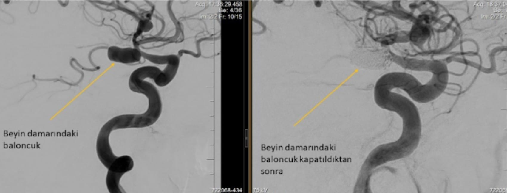
- Pre- and post-treatment images of cerebral aneurysm
Cerebral aneurysms form due to vessel wall weakening and require prompt treatment. In interventional neuroradiology, endovascular therapy is a key minimally invasive approach. Patients undergo the procedure under general anesthesia without surgical incisions.
- A catheter is inserted via the groin artery.
- It is guided to the brain through the neck vessels.
- A microcatheter is advanced into the aneurysm, and platinum coils are deployed.
- Coils block blood entry into the aneurysm, effecting treatment.
In some cases, coil embolization is supported by a stent or balloon to protect adjacent vessels. This embolization technique offers an effective alternative to open surgery, minimizing bleeding risk and accelerating recovery.
- Stent-assisted aneurysm treatment
When coils threaten the parent vessel, one or more stents (e.g., Y- or T-stenting) can be placed to preserve the parent artery and reduce recurrence risk.
- Flow-diverter stent therapy
If aneurysms are unsuitable for coiling or stent assistance, specially designed stents can treat nearly all aneurysms endovascularly.
Other Cerebral and Vascular Diseases Treated Endovascularly
Endovascular methods effectively treat various cerebrovascular diseases beyond aneurysms, including unruptured aneurysms, arteriovenous malformations (AVMs), arteriovenous fistulas (AVFs), and vessel stenoses. These techniques provide less invasive, faster recovery options:
- Treatment of hemorrhage-prone vascular bulges
- Adjunct therapy for brain and spinal tumors
- Supportive treatment for stroke and pediatric ocular tumors
Frequently Asked Questions
What should patients with cerebral aneurysm avoid?
Patients should avoid heavy exercise and lifting. Those with hypertension must monitor blood pressure regularly and adhere to antihypertensive medications. Smoking and alcohol consumption should be completely avoided to prevent aneurysm growth or rupture.
How is the onset of an aneurysm noticed?
Patients may experience double or blurred vision and eyelid drooping. These symptoms often accompany severe, persistent headaches. Nausea and vomiting can also indicate an aneurysm. Sudden muscle spasms during physical activity are another key sign. However, 95% of aneurysms remain asymptomatic and are incidentally found on brain MRIs.
Can a cerebral aneurysm resolve on its own?
No, cerebral aneurysms do not resolve spontaneously and require medical intervention. Medications cannot reduce aneurysm size or effect. Given the seriousness, physicians usually recommend surgical or endovascular treatment based on aneurysm location and characteristics.
Where does an aneurysm cause pain?
This is very rare; over 99% of headaches are not aneurysm-related. Unruptured aneurysms may be asymptomatic. When they press on surrounding structures, pain typically starts above the eye and radiates backward, often with pupil dilation, visual disturbances, or facial numbness. Any such symptoms warrant caution.
Can an aneurysm be seen on an X‑ray?
No, aneurysm detection by plain X‑ray is extremely rare. Physicians rely on detailed imaging modalities like CT angiography or MR angiography. Angiography is frequently used to determine aneurysm location and size.
Is a cerebral aneurysm visible on MRI?
MRI effectively diagnoses aneurysms larger than 4–5 mm; smaller ones are better detected with MR angiography. While MRI provides valuable information, cerebral angiography offers more detailed structural data and is often recommended for comprehensive evaluation.
Additional Resources and Documents
Flow Diverter Device Assisted Coiling Treatment for Cerebral Blister Aneurysm (PDF)
Flow Diversion of Ruptured Intracranial Aneurysm (PDF)
Unruptured Cerebral Aneurysm Risk Stratification (PDF)
Endovascular and Medical Management of Cerebral Aneurysms (PDF)

Interventional Radiology and Neuroradiology Speaclist Prof. Dr. Özgür Kılıçkesmez graduated from Cerrahpaşa Medical Faculty in 1997. He completed his specialization at Istanbul Education and Research Hospital. He received training in interventional radiology and oncology in London. He founded the interventional radiology department at Istanbul Çam and Sakura City Hospital and became a professor in 2020. He holds many international awards and certificates, has over 150 scientific publications, and has been cited more than 1500 times. He is currently working at Medicana Ataköy Hospital.


Vaka Örnekleri