A heel spur is a bony outgrowth that develops on the heel bone, often associated with plantar fascia inflammation. It results from repeated stress, leading to calcium deposits and bone formation.
The main causes of heel spurs include prolonged standing, obesity, improper footwear, and foot deformities. Athletes and individuals with flat feet are at higher risk due to continuous strain on the heel.
Symptoms include sharp heel pain, especially in the morning or after rest, along with tenderness and difficulty walking. Pain worsens with activity, limiting mobility and daily functions.
Diagnosis is confirmed through X-rays, while treatment involves lifestyle adjustments, orthotics, anti-inflammatory therapy, and physical rehabilitation. In resistant cases, shockwave therapy or surgery may be considered.
| Medical Name | Plantar Fasciitis |
| Causes | Prolonged standing, improper footwear, overweight, high-arched feet |
| Risk Factors | Obesity, standing for long periods, flat feet, unsuitable shoes |
| Common Symptoms | Sharp heel pain with the first steps in the morning, pain decreasing during the day |
| Diagnosis Methods | Physical examination, patient history, rarely X-ray or MRI |
| Complications | Chronic pain, gait abnormalities, hip/back pain |
| Treatment Methods | Rest, ice application, stretching exercises, orthotic insoles, NSAID medication, physical therapy, injections, radiofrequency, embolization |
| Surgical Necessity | Rare; considered in chronic cases not responding to conservative treatment |
| Prevention Methods | Proper footwear, regular foot exercises, weight management |
What Is a Heel Spur?
You can think of a heel spur as a defense mechanism of your body. Just like a callus forms to protect your skin in areas exposed to constant friction, your heel bone (calcaneus) reacts similarly when subjected to excessive and ongoing stress. The plantar fascia is a strong band of tissue that supports your foot like a hammock and absorbs shock with every step you take. When the area where this band attaches to the heel bone is exposed to chronic tension, small tears, and stress over many years, the body starts to deposit calcium there to strengthen the region.
This build-up does not happen overnight; it takes months or even years, eventually hardening into a bony structure—the heel spur visible on X-ray. But here’s an interesting detail: not everyone with a heel spur experiences pain. In fact, studies show that most people with a heel spur never feel any symptoms in their lifetime. These spurs are often detected incidentally when X-rays are taken for other reasons. This reminds us that the real culprit behind the pain is usually not the spur itself, but persistent inflammation of the surrounding tissue.
What Is the Relationship Between Heel Spur and Plantar Fasciitis?
These two names are often used interchangeably when talking about heel pain, because they are closely related. Plantar fasciitis refers to inflammation of the plantar fascia, that critical ligament supporting the arch of your foot. Overuse causes tiny tears and irritation in this ligament, leading to inflammation.
Their relationship is like a chicken-or-egg situation. The process typically goes as follows:
- First, the plantar fascia becomes inflamed due to overuse (Plantar Fasciitis).
- As a response to this chronic and repeated inflammation, the body begins to deposit calcium to strengthen the area.
- Over time, this deposit hardens and forms a heel spur.
In most cases, the heel spur is the result of long-standing, untreated plantar fasciitis. The bony outgrowth can further aggravate pain by putting pressure on the already inflamed tissue. That’s why treatment should focus on the stubborn inflammation rather than just the spur visible on X-ray.
What Are the Causes and Risk Factors for Heel Spurs?
Certain situations and habits can trigger or accelerate the development of a heel spur. Recognizing these risk factors is the first step toward prevention. Major risk factors include:
- Overweight or obesity
- Improper footwear
- Unsupported and flat shoes (such as ballet flats, slippers)
- Standing or walking for long periods on hard surfaces
- High-impact sports like running and jumping
- Flat feet
- High-arched feet
- Tight calf muscles and Achilles tendon
- Advancing age
- Certain rheumatic diseases
- Diabetes
What Are the Symptoms of a Heel Spur?
The pain of a heel spur is usually quite distinctive and those who experience it can easily recognize these symptoms:
- Sharp, stabbing pain in the heel with the first steps in the morning
- Similar “first step” pain after standing up following prolonged sitting
- Pain that subsides with walking but returns by the end of the day
- Localized tenderness in the underside/middle of the heel when pressed
- Avoidance of stepping on the heel and limping due to pain
- Occasional mild swelling or increased warmth in the painful area
This “first step pain” is the hallmark of both heel spur and plantar fasciitis. The reason is that the plantar fascia shortens during rest, and when you stand up, it is suddenly stretched.
How Is Heel Spur Diagnosed?
Diagnosis is mainly based on the patient’s story and physical examination, with imaging methods used for confirmation. The typical steps include:
- Listening and Examination: The first step is listening to your complaints. Details such as when the pain started, what it feels like, and when it gets worse are important. During examination, tender spots are identified, your foot structure and gait are evaluated, and calf muscle flexibility is checked. Most of the time, your symptoms and examination provide key clues.
- Imaging Methods: These confirm the diagnosis and rule out other causes (like stress fracture or cyst).
- X-ray: The gold standard for visualizing the bony outgrowth of a heel spur.
- Ultrasound (USG): Shows thickening, injury, or inflammation of the plantar fascia in detail, and is especially useful for guiding interventional treatments.
- MRI (Magnetic Resonance Imaging): Not the first choice but may be used for unclear cases or when nerve involvement is suspected.
How Does Interventional Radiology Offer Solutions for Persistent Heel Spur Pain?
If you have been suffering from heel pain for months or even years and have tried insoles, exercises, medications, even cortisone injections with no relief, interventional radiology offers modern, effective, minimally invasive alternatives to surgery. The aim of these treatments is to break the persistent cycle of inflammation—not to remove the spur itself.
One of the most innovative and successful methods is Plantar Artery Embolization (PAE). The logic is simple: chronic inflammation causes the body to create abnormal, unhealthy new blood vessels in the area. These “bad” vessels feed the inflammation and stimulate pain nerves, creating a vicious cycle.
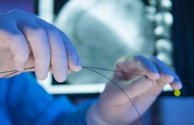
PAE works by blocking these abnormal vessels. The procedure is performed in an angiography suite, usually under local anesthesia via a tiny skin puncture at the wrist or groin. Using imaging guidance, a fine, flexible catheter is navigated to the abnormal heel vessels, and special micro-particles are injected to block their blood supply.
This cuts off the source feeding the inflammation and stops the stimulation of pain nerves, allowing the body’s natural healing process to take over. Healthy vessels are left untouched. The procedure takes about 30–60 minutes, does not require a large incision, stitches, or general anesthesia. Most patients can go home the same day and return to normal life within a few days. Especially in stubborn cases resistant to other treatments, this technique can provide long-lasting—even permanent—relief, with success rates of 80–90%.
How Can Heel Spur Formation Be Prevented?
Prevention is just as important as treatment. You can significantly reduce your risk of developing a heel spur by adopting these simple lifestyle changes:
- Maintain an ideal body weight
- Wear supportive shoes appropriate for your foot structure
- Replace old and worn-out shoes
- Avoid standing for long periods on hard surfaces
- Warm up before exercise and stretch afterwards
- Regularly stretch calf muscles and the Achilles tendon
- Wear supportive slippers at home instead of going barefoot
- Add anti-inflammatory foods (turmeric, ginger, omega-3) to your diet
When Should You See a Doctor for Heel Pain?
Not all heel pain is harmless. You should see a specialist without delay if any of the following apply to you:
- Pain has lasted for several weeks and is not improving
- Simple home remedies are not working
- Pain limits your daily life (walking, work, social activities)
- There is swelling, redness, bruising, or increased warmth in the heel
- Pain is severe enough to wake you at night or occurs at rest
- Numbness or tingling sensations accompany the pain
Heel pain is not your destiny. Especially if you have long-lasting, persistent pain that lowers your quality of life, do not hesitate to ask about modern and effective treatment options such as Plantar Artery Embolization (PAE) offered by Interventional Radiology. With accurate diagnosis and a personalized treatment plan, you can walk freely and painlessly again!

Interventional Radiology and Neuroradiology Speaclist Prof. Dr. Özgür Kılıçkesmez graduated from Cerrahpaşa Medical Faculty in 1997. He completed his specialization at Istanbul Education and Research Hospital. He received training in interventional radiology and oncology in London. He founded the interventional radiology department at Istanbul Çam and Sakura City Hospital and became a professor in 2020. He holds many international awards and certificates, has over 150 scientific publications, and has been cited more than 1500 times. He is currently working at Medicana Ataköy Hospital.


Vaka Örnekleri