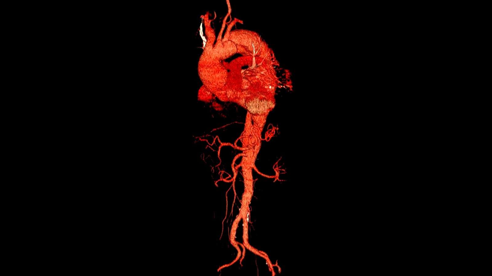
The relationship between vascular disorders and tinnitus is well established, with abnormal blood flow causing pulsatile tinnitus. Conditions such as carotid artery stenosis, arteriovenous malformations, or hypertension can generate vascular sounds perceived as ear noise.
Diagnostic evaluation includes Doppler ultrasound, CT angiography, or MR angiography. These imaging techniques help identify underlying vascular pathology responsible for tinnitus symptoms and guide treatment planning.
Management depends on addressing the vascular disorder itself. Interventions may include surgical correction, stenting, or medical therapy to restore normal blood flow and alleviate tinnitus.
Recognizing vascular causes of tinnitus is crucial, as they may indicate serious underlying diseases. Early detection ensures both symptom relief and prevention of potentially life-threatening complications.
Prof. Dr. Özgür KILIÇKESMEZ Prof. Dr. Kılıçkesmez holds the Turkish Radiology Competency Certificate, the Turkish Interventional Radiology Competency Certificate, Stroke Treatment Certification, and the European Board of Interventional Radiology (EBIR). In his academic career, he won the Siemens Radiology First Prize in 2008.
Interventional Radiology / Interventional Neuroradiology
What Is Tinnitus and How Is It Linked to Vascular Diseases?
Tinnitus is the perception of sound without an external source, usually experienced as ringing or buzzing in the ears. Tinnitus associated with vascular diseases is typically distinguished from other types by being heard in sync with the pulse. Pulsatile tinnitus occurs due to turbulent blood flow in vessels near the ears and may indicate underlying vascular issues. This type of tinnitus may be linked to the following vascular conditions:
- Arteriovenous Malformations (AVMs) and Fistulas: These involve abnormal connections between arteries and veins, causing high-pressure blood to flow abnormally and creating sounds synchronized with the pulse.
- Venous Sinus Stenosis: Narrowing of major veins that drain blood from the brain disrupts blood flow and leads to pulsatile tinnitus. This condition can cause increased intracranial pressure and result in rhythmic sounds in the ears.
- Atherosclerosis: Hardening or narrowing of the arteries results in turbulent blood flow. This can cause pulse-synchronized sounds, especially in the carotid arteries or vessels near the ear.
The diagnostic process for pulsatile tinnitus generally includes advanced imaging methods such as MRA and CTA. Treatment focuses on addressing the underlying vascular issues and aims to improve the patient’s quality of life.
How Does Cardiovascular Health Affect Tinnitus?
Cardiovascular health has a significant impact on the auditory system and is one of the main factors that can trigger tinnitus. Arterial narrowing due to conditions such as atherosclerosis can lead to permanent damage to hearing-related structures like the cochlea. These vascular issues may prevent adequate oxygen from reaching the delicate hair cells in the cochlea, causing tinnitus. Especially cardiovascular disorders such as hypertension increase the risk of tinnitus by affecting blood flow. Many studies have shown this link, with individuals who have high blood pressure being at greater risk of experiencing tinnitus symptoms.
The effects of cardiovascular diseases such as arterial hardening and narrowing on ear health have been examined through various mechanisms. These effects are listed below:
- Decreased blood flow: Diseases such as atherosclerosis and hypertension make it difficult for blood to reach auditory structures like the cochlea.
- Permanent cell damage: Lack of oxygen can cause irreversible damage to hair cells in the cochlea.
- Increased risk of pulsatile tinnitus: Blood flow irregularities caused by cardiovascular diseases can trigger the form of tinnitus felt in sync with the heartbeat.
*We recommend filling out all fields so we can respond in the best possible way.
Can Hypertension Cause Tinnitus?
Hypertension can cause a type of tinnitus known as pulsatile tinnitus, which usually manifests as sounds synchronized with the heartbeat. High blood pressure, especially by increasing pressure on vessels around the ear, leads to turbulent blood flow. This situation, along with increased pressure on vessel walls, can affect inner ear structures and cause the constant perception of phantom sounds. Since hypertension affects the speed and dynamics of blood flow, it can increase the intensity of tinnitus symptoms.
Effectively controlling hypertension can alleviate pulsatile tinnitus symptoms and improve quality of life. The following treatment and lifestyle measures are recommended:
- Medication: Blood pressure-lowering drugs such as beta blockers, ACE inhibitors, and diuretics can reduce pressure on vessel walls and control turbulence.
- Lifestyle Changes: Reducing sodium intake, regular physical exercise, and practicing stress management are important for managing hypertension and related tinnitus.
- Avoiding Alcohol and Smoking: These substances can raise blood pressure, contributing to hypertension and thus tinnitus.
How Do Atherosclerosis and Carotid Artery Disease Affect Tinnitus?
Atherosclerosis and carotid artery disease play a significant role in the development of pulse-synchronized tinnitus. Plaques that accumulate in the carotid arteries during atherosclerosis cause these vessels to narrow and harden. This narrowing leads to turbulent blood flow, especially near the middle and inner ear, and patients hear a “whirring” sound synchronized with their pulse. This sound occurs because blood flow becomes irregular in narrowed arteries, and the ear starts to perceive these flow disturbances.
Over time, impaired blood flow to the inner ear can affect cochlear microcirculation and lead to hearing problems. Decreased blood flow causes ischemia in ear structures, contributing to the onset of tinnitus. Additionally, increased vascular resistance in narrowed arteries not only worsens tinnitus but also raises the risk of stroke. Arterial stiffening associated with atherosclerosis further strengthens the vascular causes of tinnitus. This arterial rigidity reduces vessel elasticity, further impedes blood flow, and increases tinnitus severity.
Major risks include:
- Impaired blood flow to the inner ear: Affects hearing function and increases tinnitus severity.
- Increased vascular resistance: Can lead to serious health issues such as stroke.
- Arterial hardening: Disrupts blood flow dynamics and creates broader cardiovascular risks.
How Do Vascular Malformations Cause Tinnitus?
Vascular malformations such as arteriovenous malformations (AVMs) and aneurysms can affect the dynamics of blood flow around the ear and lead to pulsatile tinnitus. Pulsatile tinnitus presents as a rhythmic sound synchronized with the heartbeat, and turbulent blood flow can trigger this sound. Especially arteriovenous malformations (AVMs) near the ear disrupt normal blood flow by forming direct connections between arteries and veins. This creates abnormal blood flow near the ear, resulting in the perception of vibratory sounds.
Aneurysms occur in weak and bulging areas of arteries. When these structures develop in vessels close to the ear, such as the carotid artery, they can cause symptoms of pulsatile tinnitus. Turbulent blood flow along aneurysms causes the ear to perceive vibrating sounds.
Both types of vascular malformations can cause serious health problems. Since they increase risks such as sudden rupture or bleeding, early diagnosis and management are very important. When pulsatile tinnitus is detected, if additional symptoms such as headaches or neurological findings are present, urgent intervention is required. Early diagnosis is vital, especially for preventing complications such as intracranial hemorrhage or stroke.
Neuroimaging studies are generally requested to confirm the diagnosis:
- Magnetic resonance angiography (MRA)
- Computed tomography angiography (CTA)
What Diagnostic and Treatment Options Are Available for Vascular-Origin Tinnitus?
Various methods are used for the diagnosis and treatment of vascular-origin tinnitus. First, imaging techniques play a critical role in diagnosing this condition. Magnetic Resonance Angiography (MRA) provides detailed images of both arterial and venous structures and is used to detect stenosis or arteriovenous malformations in blood vessels. Additionally, Computed Tomography Angiography (CTA) is also widely preferred and offers effective results, especially in identifying arterial abnormalities.
Among other diagnostic tools, cerebral angiography stands out. This method helps detect vascular problems with detailed vascular imaging in cases where other non-invasive tests are inconclusive. Additionally, Carotid Duplex Ultrasound is used to measure blood flow, especially for the detection of arterial stenosis. In cases of non-vascular causes, additional evaluations can be performed with high-resolution temporal bone CT or brain MRI.
During the treatment process, lifestyle changes focus on reducing cardiovascular risks and include the following measures:
- Keeping blood pressure under control
- Lowering cholesterol levels
- Managing conditions such as diabetes
Medical treatments include antihypertensive drugs and statins. These drugs improve vascular health and may alleviate tinnitus. Surgical and interventional procedures have expanded treatment options, and endovascular stenting and embolization can be preferred in such cases.
In cases of venous sinus stenosis, stenting is performed, and in venous aneurysms, coiling or stenting may be used. In the presence of vascular malformation or fistula, endovascular embolization by angiographic methods will eliminate disturbing sounds and prevent possible brain hemorrhages.

Interventional Radiology and Neuroradiology Speaclist Prof. Dr. Özgür Kılıçkesmez graduated from Cerrahpaşa Medical Faculty in 1997. He completed his specialization at Istanbul Education and Research Hospital. He received training in interventional radiology and oncology in London. He founded the interventional radiology department at Istanbul Çam and Sakura City Hospital and became a professor in 2020. He holds many international awards and certificates, has over 150 scientific publications, and has been cited more than 1500 times. He is currently working at Medicana Ataköy Hospital.


Vaka Örnekleri