


A cyst is an abnormal sac-like structure in the body filled with fluid, semi-solid material, or gas. Cysts are usually benign and can occur under the skin, in organs, or within tissues. Symptoms may vary depending on the location, size, and type of the cyst.
Causes of cysts include infections, genetic predisposition, chronic inflammation, or blocked ducts. Depending on the cause, some cysts may shrink and disappear on their own, while others may require intervention.
The most common types of cysts include epidermoid cysts, ovarian cysts, ganglion cysts, and kidney cysts. Each type of cyst carries symptoms and risks specific to the region where it forms, making the diagnostic process important.
Cyst symptoms generally include pain, swelling, limited movement, or functional impairment due to pressure. While some cysts may be asymptomatic, others can lead to serious complications. Imaging methods such as ultrasound, MRI, or CT are used for diagnosis.
What Is a Cyst and Why Does It Form in Our Body?
To understand the mechanism of cyst formation, it is necessary to examine the body’s defense and repair systems. The body tends to isolate a material that it perceives as foreign or cannot manage, in order to prevent it from harming other healthy tissues. This isolation process occurs by forming a wall around that material. If this wall is a membrane made of real cells, it is called a true cyst; if it is surrounded only by inflammatory tissue, it is called a pseudocyst. However, from the patient’s perspective, the perceived result is generally similar.
The underlying causes of cyst formation vary greatly from person to person. One of the most common causes is the blockage of fluid channels in the body. Our body continuously produces various fluids and secretions. For example, the sebaceous glands in our skin produce sebum to keep the skin moist. If the channel through which this fluid reaches the skin becomes blocked, production continues internally but there is no external drainage. In this case, the accumulated fluid expands backward, forming a cyst.
Another important cause is infections and parasites. One of the best examples is hydatid cyst, which is frequently encountered in our country and is a serious public health problem. Parasites transmitted through unwashed vegetables or infected animals settle in organs such as the liver or lungs and form fluid-filled cysts. This is the body’s attempt to isolate the parasite.
Sometimes our genetic inheritance also plays a role. Certain inherited genetic traits can predispose individuals to the formation of multiple cysts, as seen in the kidneys. Additionally, remnants of tissue that should disappear during fetal development may remain in the body, leading to the formation of structures known as dermoid cysts later in life. Trauma should not be overlooked either; impacts or strain on joints can cause joint fluid to herniate outward and form a cystic structure.
What Are the Symptoms of a Cyst and How Does It Manifest?
One of the most common questions our patients ask is how they can tell if they have a cyst in their body. Cysts are usually insidious structures. When they are small, they produce almost no symptoms and can remain unnoticed for years. However, as the cyst grows, it begins to make itself felt by stretching the capsule of the organ it is located in or by exerting pressure on neighboring organs.
The severity and nature of symptoms vary depending on the location of the cyst. Generally, the physical effect caused by a cyst is a feeling of fullness and pressure. If the cyst is close to the surface, the patient may complain of a palpable lump. In deeper organs, it usually manifests through dysfunction or pain.
As the nature of the cyst changes, the symptoms also change. For example, a simple cyst filled with clear fluid may remain silent, but if bleeding occurs inside it or if it becomes infected, the condition can suddenly become more severe. The pain intensifies, there may be an increase in temperature in the area, and the patient may feel fatigued.
Common complaints associated with cysts include:
- Palpable mass
- Localized swelling
- Pain
- Feeling of fullness
- Pressure sensation
- Limited movement
- Loss of function
- Redness
What Are the Symptoms of Liver Cysts and What Risks Do They Carry?
The liver functions like a factory of our body and is one of the internal organs where cysts are most commonly seen. When evaluating liver cysts, we divide them into two main groups: simple cysts and parasitic cysts. Simple cysts are usually benign and often congenital. Although they occupy space within the liver tissue, they generally do not impair vital functions such as blood filtration or bile production. However, when they exceed 10 centimeters in size or are numerous, they begin to alter intra-abdominal balance.
A large liver cyst can stretch the outer membrane of the liver, causing a constant, dull pain under the right rib. This pain may sometimes radiate to the right shoulder. More importantly, it can put pressure on the stomach, which is located adjacent to the liver. When the cyst compresses the stomach, the patient may feel full even after eating a small amount. This is called early satiety. Indigestion, bloating, and gas complaints may accompany this.
Hydatid cyst (parasitic cyst), on the other hand, requires much more careful management. These cysts are alive and tend to grow. The biggest risk is rupture. If the cyst ruptures due to trauma or spontaneously, its contents spread into the abdominal cavity. Since this fluid is foreign to the body, it can cause a severe allergic reaction or even shock.
The main problems that liver cysts can cause include:
- Abdominal pain
- Indigestion
- Early satiety
- Nausea
- Vomiting
- Jaundice
- Fever
- Shortness of breath
What Complaints Do Kidney Cysts Cause and What Is Their Relationship with Blood Pressure?
Kidney cysts are a condition that is almost as common as graying hair, especially in individuals over the age of 50. Most are called “simple cortical cysts” and are harmless. However, when the cyst grows or becomes complex in structure, it begins to send interesting signals in the body.
The most typical symptom of kidney cysts is what we call “flank pain,” felt on the side of the back, at the end of the ribs. This pain can sometimes be confused with a herniated disc or muscle pain. However, the most surprising effect of a cyst is on blood pressure. The kidneys are the main center that regulates the body’s blood pressure. When a large cyst puts pressure on the kidney tissue or vessels, the kidney perceives that it is not receiving enough blood and releases a hormone called “renin.” This hormone constricts the vessels and raises blood pressure. In other words, in a patient with unexplained, drug-resistant hypertension, the underlying cause may sometimes be a seemingly innocent kidney cyst.
In addition, if the cyst presses on the urinary tract, it can obstruct urine flow and cause swelling in the kidney (hydronephrosis). This increases the risk of infection. If bleeding occurs inside the cyst or if it opens into the urinary tract, the patient may see blood in the urine, which is one of the most alarming symptoms for our patients.
The main symptoms seen in kidney cysts include:
- Flank pain
- Back pain
- High blood pressure
- Bloody urine
- Frequent urination
- Burning during urination
- Fever
- Abdominal swelling
Can a Thyroid Cyst Cause Hoarseness?
The thyroid gland, located at the front of our neck, is highly prone to nodule formation, and a significant portion of these nodules are cystic, meaning fluid-filled structures. Thyroid cysts are generally low-risk in terms of malignancy (cancer), but due to their location, they are strategically positioned. Directly behind them are the trachea and esophagus, and on the sides are the carotid arteries and the nerves leading to the vocal cords:
Patients usually present after noticing an asymmetry in their neck or a soft swelling they can feel with their hands. While the cyst may initially appear to be only a cosmetic issue when small, as it grows it begins to compress the structures behind it. The patient may feel as if something is stuck in their throat when swallowing. They may feel as though there is a constant lump in their throat and feel the need to clear it.
Larger cysts can press on the airway, causing shortness of breath, especially when lying down, and noisy breathing. The most critical symptom is hoarseness. If the cyst compresses the nerve that controls the vocal cords, it can cause voice cracking, deepening, or hoarseness. Additionally, if sudden bleeding occurs inside the cyst, it can cause sudden severe pain in the neck and rapid enlargement of the swelling.
The main discomforts caused by thyroid cysts include:
- Swelling in the neck
- Difficulty swallowing
- Sensation of something stuck in the throat
- Hoarseness
- Deepening of the voice
- Shortness of breath
- Neck pain
- Cough
What Is a Baker’s Cyst and Why Does Swelling Occur Behind the Knee?
If you feel tension, fullness, or an egg-sized swelling in the hollow area behind your knee (popliteal fossa), it is most likely called a Baker’s cyst. This cyst is actually a response to a problem in the knee joint. There is a special fluid inside the knee joint that provides lubrication. In conditions such as meniscus tears, osteoarthritis, or rheumatism, the body produces this fluid in excess to protect the knee. As pressure increases inside the joint, this excess fluid herniates outward through a weak point behind the knee, forming a sac.
Patients often describe this condition as “I can’t fully bend my knee, something feels stuck behind it” or “When I straighten my leg completely, I feel tension behind it.” The swelling usually increases with standing or walking and may decrease somewhat with rest.
The most dramatic moment of a Baker’s cyst is when it ruptures. If the cyst wall tears, the thick fluid inside leaks between the calf muscles. This causes severe pain, redness, and swelling in the calf. This condition is often confused with deep vein thrombosis (DVT). Differentiating between the two is of vital importance because their treatments are completely opposite.
Typical symptoms of a Baker’s cyst include:
- Swelling behind the knee
- Tension behind the knee
- Limited movement
- Knee pain
- Calf pain
- Swelling in the leg
- Redness
How Is a Cyst Diagnosed and What Methods Are Used?
In a patient presenting with suspected cyst, it is not possible to fully understand the structure of the cyst with only a physical examination. Is the content of the cyst fluid or solid? Is its wall thick? Is there bleeding inside? To answer these questions, we use advanced imaging technologies. The more accurate the diagnosis, the more successful the treatment.
The first and most commonly used method is Ultrasonography (USG). Since it does not involve radiation, it can be safely used even in pregnant women and children. Ultrasound works using sound waves and is excellent at identifying fluid-filled structures. While a cyst appears completely black (anechoic) on the screen, solid masses appear gray or white. This allows us to largely distinguish whether the cyst is a simple fluid sac or a structure suspicious for a tumor.
If more detailed mapping is required, Computed Tomography (CT) is used. CT helps us understand the relationship of the cyst with surrounding vessels, detect calcifications, and plan surgical or interventional procedures.
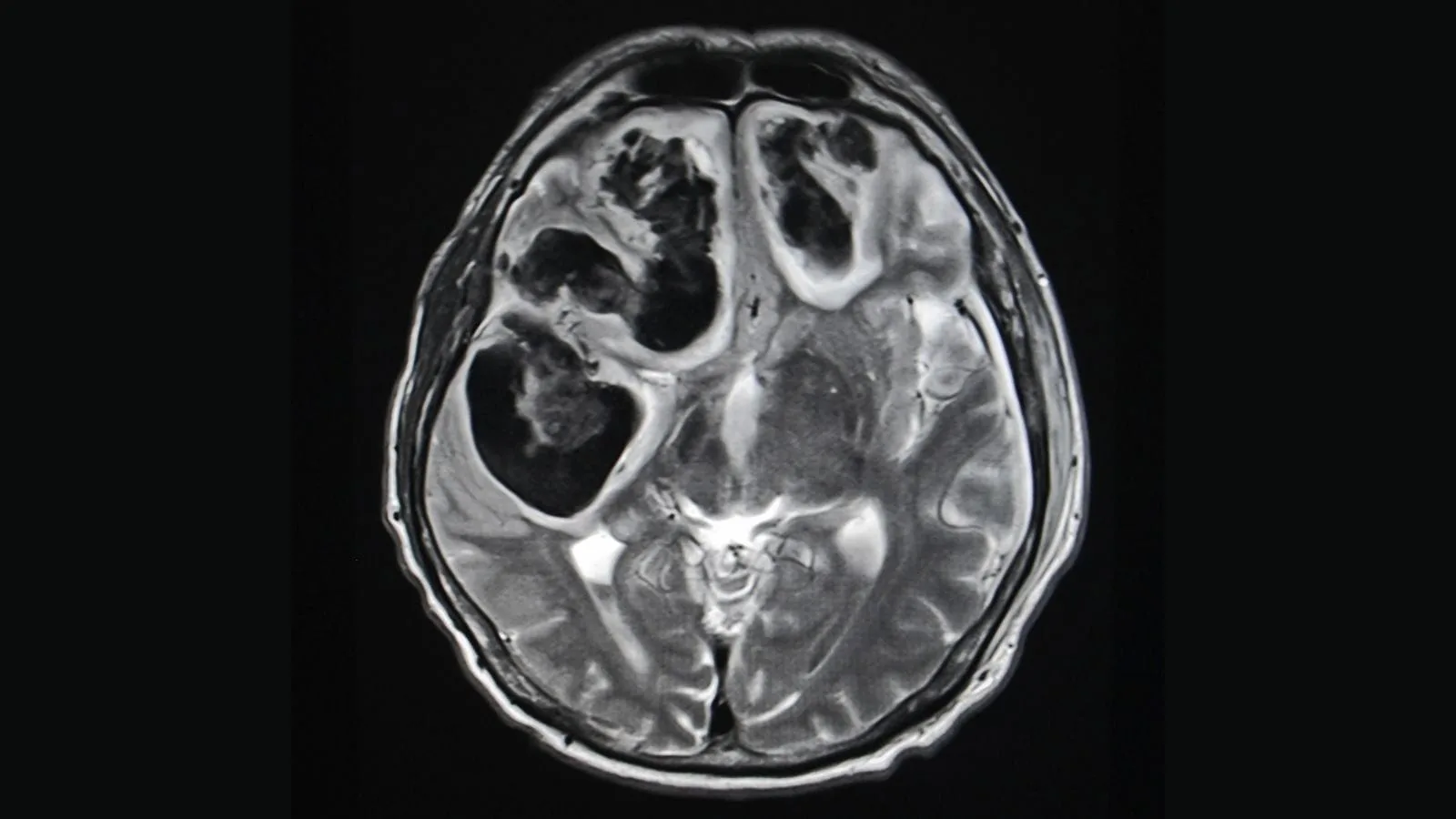
The method that shows the internal structure of the cyst in the greatest detail is Magnetic Resonance Imaging (MRI). MRI is indispensable, especially in suspected hydatid cysts of the liver or cysts involving the brain. It allows us to determine whether the internal membranes of the cyst are separated and whether there is a connection with the bile ducts.
Methods used in the diagnostic process include:
- Physical examination
- Ultrasonography
- Doppler ultrasonography
- Computed tomography
- Magnetic resonance imaging
- Blood tests
Is Non-Surgical Cyst Treatment Possible and What Is Interventional Radiology?
For many years, the only treatment for cysts was surgery. Patients would undergo anesthesia, incisions would be made in their bodies, and the cyst would be removed. However, developments in medical technology have brought forward a field called “Interventional Radiology,” which can be considered the seeing eye of modern medicine. This specialty performs treatments under the guidance of imaging devices (such as ultrasound and tomography), entering through needle holes without making large incisions in the body.
The best news we can give our patients is this: Yes, today a large majority of cysts can be treated without surgery, anesthesia, or sutures. The aim of these methods is to intervene only on the problematic cyst without disrupting the anatomical integrity of the patient. These approaches, which we can also call “targeted treatment,” aim to restore health without confining the patient to a hospital bed or disconnecting them from daily life.
How Is Needle Aspiration and Alcohol Treatment of a Cyst Performed?
In simple cysts, the first solution that comes to patients’ minds is “draining the fluid with a needle.” Although it may seem logical, this procedure alone is unfortunately not sufficient. As long as the wall that forms the cyst remains in place, the cyst fluid will be produced again and the cyst will return in a short time, sometimes even larger than before.
To prevent this recurrence, we apply a method called “sclerotherapy.” The procedure is performed under local anesthesia while communicating with the patient. By visualizing the cyst on the ultrasound screen, we enter it with a thin needle. First, we completely drain the fluid inside the cyst. The cyst becomes like a deflated balloon. However, our job is not finished. To destroy the lining layer (epithelium) that produces fluid in the cyst wall, we inject an “adhesive/drying” substance into the cyst. The most commonly used substance for this procedure is pure sterile alcohol (ethanol). Alcohol kills the cells in the cyst wall and causes the walls to adhere to each other. After waiting approximately 15–20 minutes, we aspirate the alcohol. Thus, the cyst cavity is permanently sealed.
What Is the PAIR Method in Hydatid Cyst Treatment?
The PAIR method used in the treatment of parasitic cysts (hydatid cysts) is a powerful alternative to surgery. PAIR is an acronym formed from English words: Puncture, Aspiration, Injection, and Re-aspiration. The principle is similar, but the procedure must be performed much more carefully.
Under ultrasound guidance, the cyst is entered and the pressurized fluid inside is drained in a controlled manner. Once the pressure inside the cyst decreases, special agents that kill the parasite (usually high-concentration saline or alcohol) are injected. This medication is left inside the cyst to ensure the complete destruction of the parasite’s active layer and larvae. In the final stage, the injected substance is re-aspirated. This method significantly reduces the risk of rupture and spread into the abdomen compared to open surgery. Additionally, the patient’s recovery time is measured in hours rather than days.
What Special Methods Are Used for Thyroid and Baker’s Cysts?
The “Percutaneous Ethanol Ablation” (PEA) method we apply in cystic nodules of the thyroid gland has significantly reduced the need for thyroid surgery. Since part or all of the gland is removed during thyroid surgery, the patient may need to use hormone medication for life. However, in ablation therapy, only the cyst is targeted, and the healthy thyroid tissue is preserved. Alcohol is injected into the cyst to shrink the nodule. There are no concerns such as hoarseness risk after the procedure or surgical scars.
In Baker’s cysts, the approach is both to drain the cyst and to suppress inflammation. Under ultrasound guidance, the cyst fluid behind the knee is drained. However, to prevent the cyst from refilling and to reduce edema in the area, a cortisone (steroid) injection is usually administered into the cyst. This procedure quickly relieves the patient from pain and the sensation of tension.
What Are the Advantages of Interventional Radiology?
Interventional radiological methods offer many advantages in terms of comfort and safety compared to classical surgery. These methods, which align with the principle of modern medicine “do the least harm to the patient,” maximize patient satisfaction.
The main advantages of these methods include:
- Does not require general anesthesia.
- Performed with only local anesthesia.
- No incision or suture marks on the skin.
- Very low risk of infection.
- Very short recovery time.
- The patient can return home the same day.
- Healthy tissues are preserved.
- Less painful.
Can a Cyst Recur After Treatment and How Should Follow-Up Be?
As with any medical procedure, follow-up is very important in cyst treatments. We do not expect the cyst to disappear instantly after an interventional procedure; it is a process. The body gradually cleans and shrinks the treated and deflated cyst wall over time.
In kidney and liver cysts, we perform ultrasound follow-ups at the 1st, 3rd, and 6th months after the procedure. In these checks, we confirm that the cyst cavity has closed and that fluid has not reaccumulated. Success rates are quite high, but in rare cases, the cyst may refill; in such situations, the procedure can be easily repeated. In thyroid cysts, shrinkage continues over months. While a 50% reduction is observed in the first months, by the end of one year, the cyst may become almost invisible.
Frequently Asked Questions
What is a cyst and how does it develop in different parts of the body?
A cyst is a closed sac-like structure that may contain fluid, air, pus, or other material. Cysts can develop in various tissues and organs due to blockage, infection, inflammation, injury, or other underlying conditions.
What are the most common symptoms of a cyst depending on its location?
Symptoms vary by location and size but may include a visible lump, swelling, pain, tenderness, pressure, or discomfort. Some cysts cause no symptoms and are discovered incidentally during medical examinations.
Can a cyst cause pain even if it is not cancerous?
Yes, many benign cysts can cause pain if they become inflamed, infected, enlarged, or press on nearby nerves and tissues. The severity of symptoms often depends on the cyst’s location and size.
What causes cysts to form in the skin, organs, or glands?
Cysts may develop due to blocked ducts, infections, chronic inflammation, genetic conditions, hormonal changes, tissue injury, or abnormal cell growth. The exact cause depends on the type and location of the cyst.
How can doctors determine whether a cyst is benign or potentially serious?
Evaluation may include a physical examination, ultrasound, CT scan, MRI, or biopsy. These tests help assess the cyst’s characteristics and determine whether further investigation or treatment is necessary.
Can a cyst disappear on its own without medical treatment?
Some cysts may shrink or resolve spontaneously, particularly small and uncomplicated ones. Others can persist, enlarge, recur, or require treatment depending on their cause, location, and associated symptoms.
What symptoms suggest that a cyst should be evaluated by a healthcare professional?
Medical evaluation is recommended for rapidly growing cysts, persistent pain, redness, fever, drainage, bleeding, difficulty swallowing, breathing problems, or any lump with unusual or changing characteristics.
What treatment options are available for symptomatic or large cysts?
Treatment may include observation, medication, drainage, aspiration, or surgical removal. The most appropriate approach depends on the cyst type, symptoms, risk of recurrence, and underlying cause.
Can cysts recur after they have been drained or treated?
Yes, some cysts can return if the cyst wall remains intact or if the underlying cause persists. Surgical removal of the entire cyst may reduce the likelihood of recurrence in certain situations.
Are all cysts harmless or can some be associated with cancer?
Most cysts are benign and noncancerous. However, some cystic lesions may require further evaluation to exclude malignancy, especially if they have suspicious imaging features or unusual clinical characteristics.

Prof. Dr. Özgür Kılıçkesmez is one of Türkiye’s leading interventional radiology and neuroradiology specialists, focusing on minimally invasive endovascular treatments. His primary areas of expertise include aneurysm and stroke treatments, embolization procedures, non-surgical treatments of thyroid and parathyroid diseases, interventional oncology, and peripheral vascular interventions. He currently serves as the Head of the Interventional Radiology and Neuroradiology Department at Istanbul Memorial Göztepe Hospital.
Graduating from Istanbul University Cerrahpaşa Faculty of Medicine in 1997, Prof. Dr. Kılıçkesmez received advanced training in interventional radiology and oncology as a Clinical Fellow at London Guy’s & St Thomas’ and King’s College Hospital between 2012 and 2013. He earned the title of Professor in 2020 and, in the same year, became the Founding Interventional Radiologist at SBÜ Çam and Sakura City Hospital. He also holds the EBIR (European Board of Interventional Radiology) certification.
Prof. Dr. Kılıçkesmez has 94 international and 40 national publications and over 2500 citations. He serves as a reviewer for prestigious journals such as the American Journal of Roentgenology (AJR) and Diagnostic & Interventional Radiology. With his clinical and academic expertise, he is a nationally and internationally recognized authority in stroke treatment, brain aneurysm, embolization, tumor ablation, and minimally invasive treatments of thyroid/parathyroid diseases.




Vaka Örnekleri